
Figure 20.
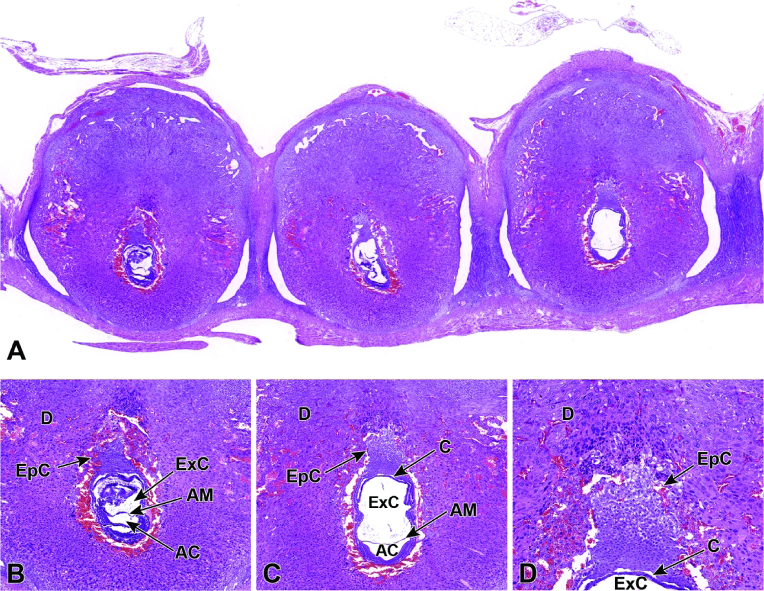
Representative images of a uterine horn showing three implantation sites at E7.5. Figure A: Viewing multiple implantation sites at once can help correctly age and identify formed placental (and embryonic) structures, as conceptuses of the same chronological age can differ by up to 24 hours when the stage of development is judged by structural features. Note that the orientation differs for placentas and embryos in adjacent implantation sites, with only some showing an ideal view. Figures B and C: The allantois has formed and has begun to cross the exocoelomic cavity (ExC) toward the chorionic plate (C). During this allantois spanning process, the proamniotic cavity and the exocoelomic cavity become distinct from one another, forming the amniotic cavity (AC) and the amnion (AM). Figure D: There is invasion of the ectoplacental cone (EpC) into the maternal decidua (D) in preparation for labyrinth induction.