
Figure 23.
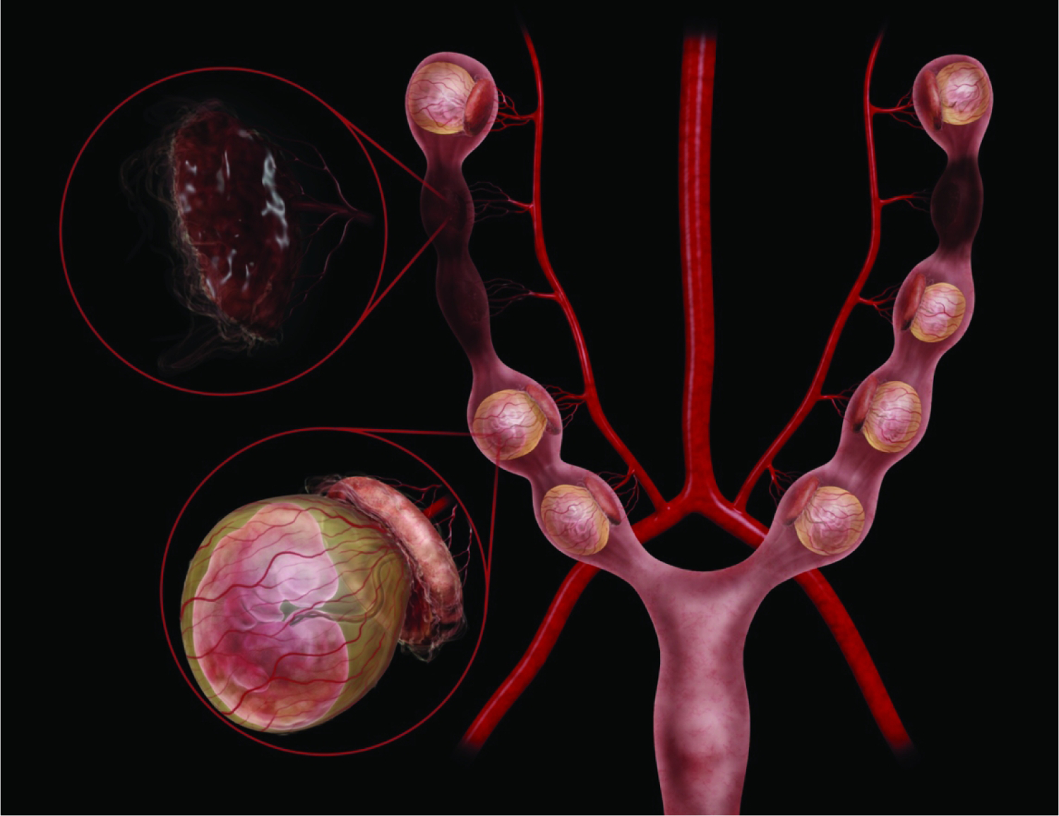
Diagrammatic representation of the mouse right and left uterine horns at E9.5. Illustrations here show both healthy, intact embryos and placentas (lower left magnification circle, with the definitive placenta capping the yolk sac-enveloped embryo) as well as some resorptions (upper left magnification circle, with collapsed placental residue). Within a single litter there can be both viable and non-viable conceptuses (i.e., embryo and placenta pairs), and among the viable offspring their developmental ages can be offset by as much as 24 hours. Embryo positioning within the yolk sac, in relationship to the placenta, can vary from embryo to embryo. The uterine vascular supply is from the aorta by way of the left and right uterine arteries, which then branch to form the arcuate and radial arteries that supply each developing embryo. 180×139mm (800 × 800 DPI)