
Figure 27.
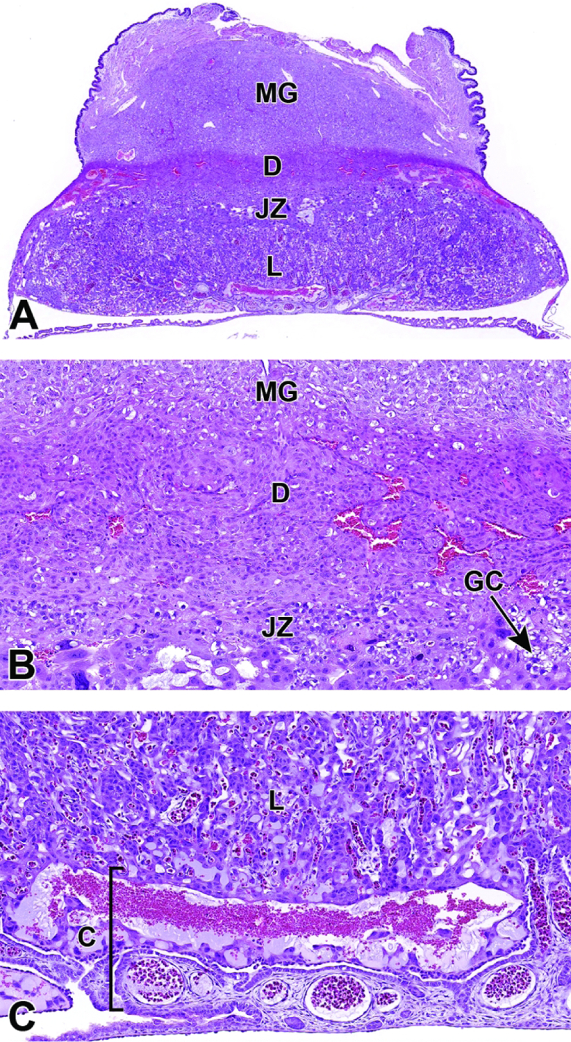
Representative images of a placental cross-section at E12.5 showing the fully functional definitive placenta with adjacent metrial gland (MG). Figure A: Low magnification of a placenta cross-section highlighting the various placental layers. Figure B: Higher magnification of the maternal decidua (D) and junctional zone (JZ). The junctional zone is composed primarily of glycogen cells (GC) and spongiotrophoblast cells that can form branching columns that extend into the labyrinth. Figure C: Higher magnification of the labyrinth (L) and chorionic plate (C). The highly vascular chorionic plate is the basal site for attachment of the umbilical cord vessels. At this stage the labyrinth makes up half of the entire placental weight as it fills with embryonic and maternal derived blood.