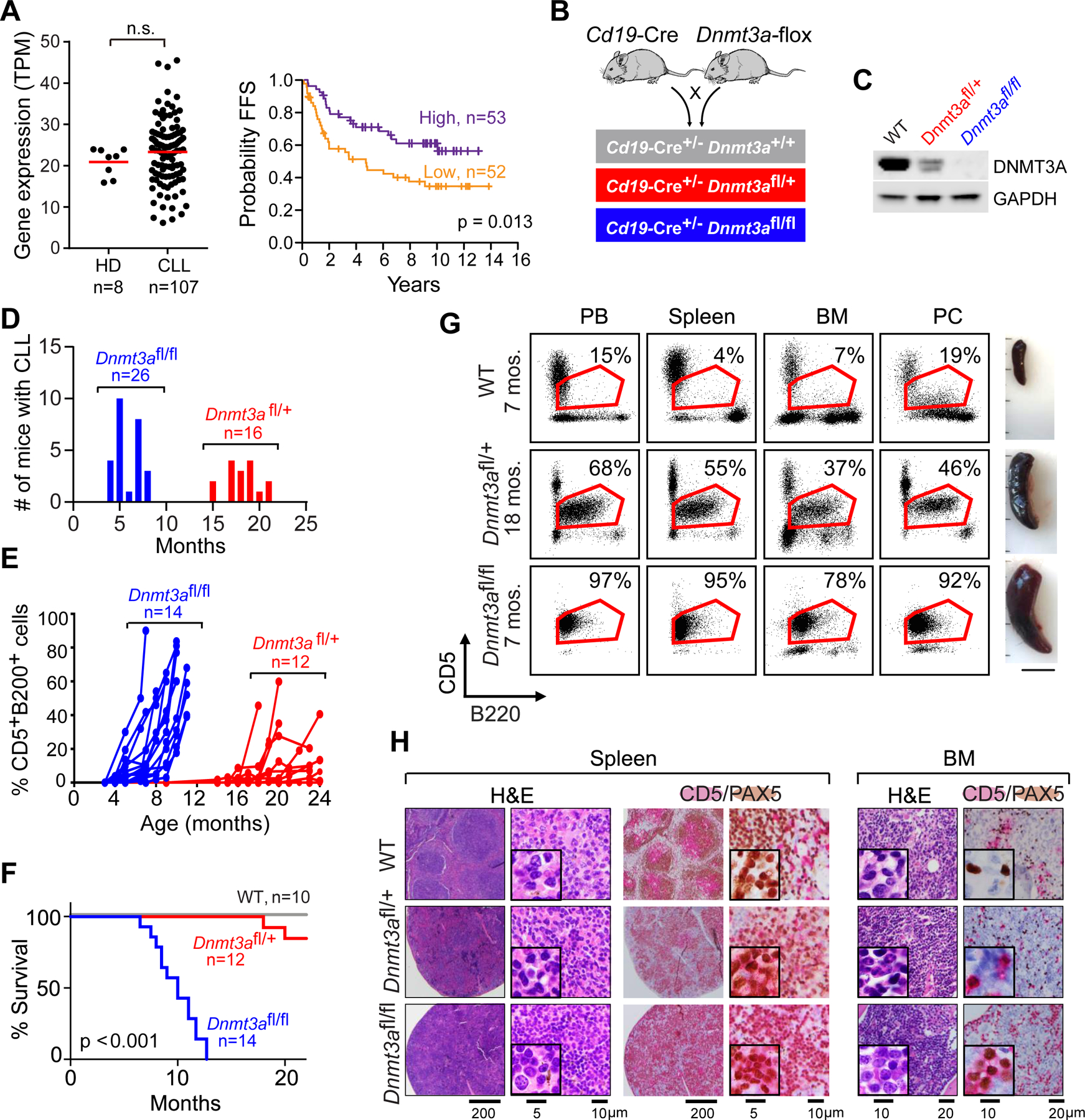
Figure 1: B cell-restricted deletion of Dnmt3a leads to CLL.

A, DNMT3A expression in human normal B cells (HD) and CLLs. Failure-free survival (FFS) from the time of sequencing sample to 1st treatment/progression or death in the DNMT3A high or low group is shown. Samples were stratified by median DNMT3A expression; there were 2 dropouts. B, Schema of the breeding strategy. C, Representative western-blots of DNMT3A expression in splenic B cells across the different genotypes (n=3). D, Age of disease onset in Dnmt3afl/+ and Dnmt3afl/fl mice. E, Detection of B220+CD5+ cells in peripheral blood over time. F, Overall survival of WT, Dnmt3afl/+ and Dnmt3afl/fl mice over time. G, Representative flow cytometry analysis of B220+ CD5+ cells within peripheral blood (PB), spleen, bone marrow (BM) and peritoneal cavity (PC) of WT, Dnmt3afl/+ and Dnmt3afl/fl CLL mice and images of the spleens. H, Representative immunohistochemical staining of CD5+(red) PAX5+ (brown) of spleen and bone marrow (BM) sections from WT and Dnmt3afl/fl CLL mice.
