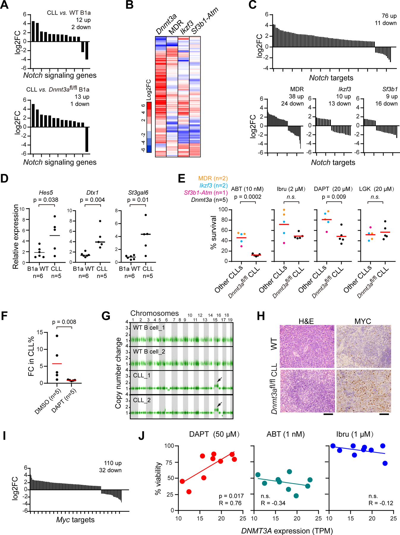
Figure 5: Activation of Notch-Myc signaling in Dnmt3afl/fl CLLs.

A, Changes in expression of significantly altered Notch signaling genes in Dnmt3afl/fl CLLs versus WT B1a cells. B, Heatmap of change in expression of Notch signaling genes in different mouse models. C, Changes in significantly altered Notch target genes in different CLL mouse models. D, qPCR analysis of expression of well-known Notch target genes in different cells. E, Sensitivity of different CLL cells with various driver mutations to different drugs, including the BCL2 inhibitor ABT-199 (venetoclax, ABT), BTK inhibitor Ibrutinib (Ibru), the Notch signaling inhibitor DAPT, and Wnt signaling inhibitor LGK-974 (LGK). F, Fold-change (day 14 vs. day 0) of the percentage of circulating CLL cells of transplanted mice treated with DMSO or 30mg/kg DAPT. G, Copy number analysis of WT B cells or Dnmt3afl/fl CLL (n=2). Arrows: Amplification of Chr 15. H, IHC analysis of Myc in spleen sections of Dnmt3afl/fl CLLs versus WT B1a cells. I, Change in expression of Myc targets in Dnmt3afl/fl CLLs versus WT B1a cells. J, The correlation between DNMT3A expression and response to different drugs in human primary CLLs (n=9) in vivo, measured by CellTiterGlo.
