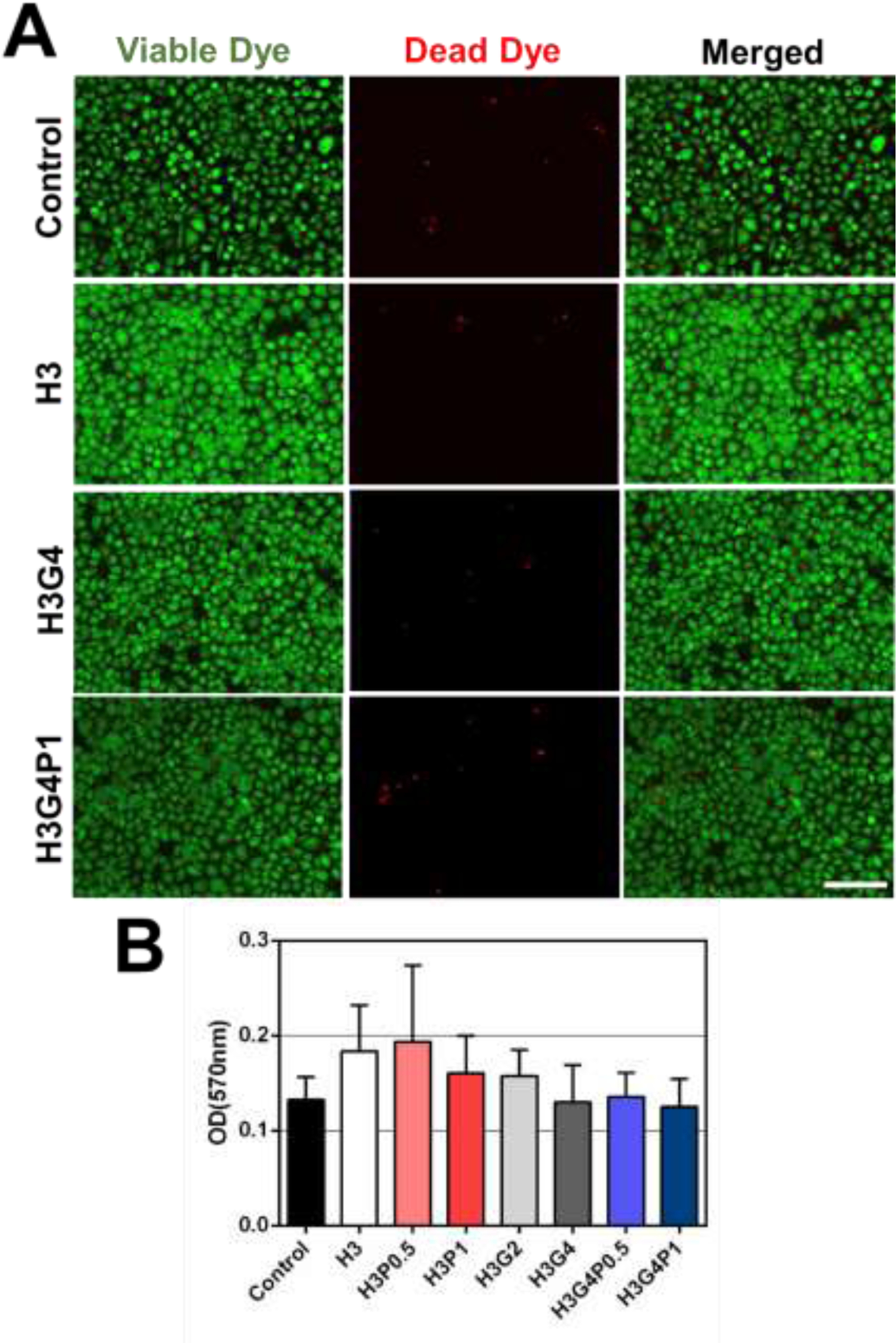
Figure 4. In vitro cytocompatibility of the hydrogels after photocrosslinking.

(A) Microscopic images of human corneal epithelial cells (HCECs) incubated for 24 h with culture medium or fluid extracts of photocrosslinked hydrogels prepared with different concentrations of HAGM, GelMA and PEGDA and then stained with calcein-AM (green - viable dye) and propidium iodide (red - dead dye) (n=3 per group), scale bar = 100 μm. (B) Cell viability measured by PrestoBlue assay after 3-days incubation with hydrogels prepared with different concentrations of HAGM, GelMA and PEGDA. Data is represented as mean ± SD (n=3 per group).
