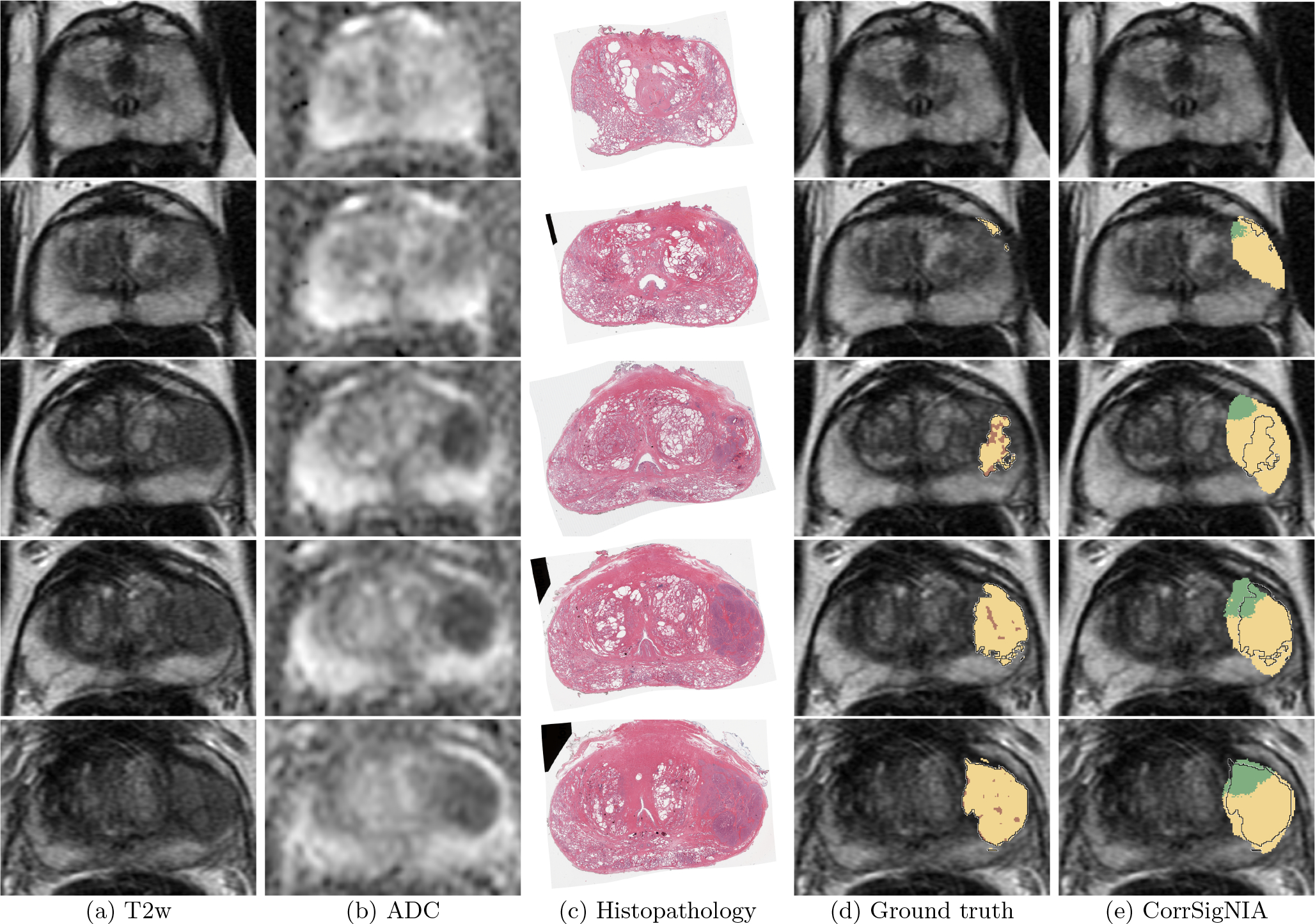
Fig. 5:

Detection and localization of aggressive cancer in a patient in cohort C1 shown from apex (top row) to base (bottom row). Registered (a) T2w image, (b) ADC image, (c) histopathology image, (d) T2w image overlaid with ground truth labels: cancer from expert pathologist (black outline), aggressive cancer (yellow) and indolent cancer (green) histologic grading from (Ryu et al., 2019), pixels within pathologist outline without automated histologic grade labels shown in brown, (e) T2w image overlaid with predicted labels from CorrSigNIA: predicted aggressive cancer (yellow) and predicted indolent cancer (green), black outline represents ground truth pathologist cancer outline.
