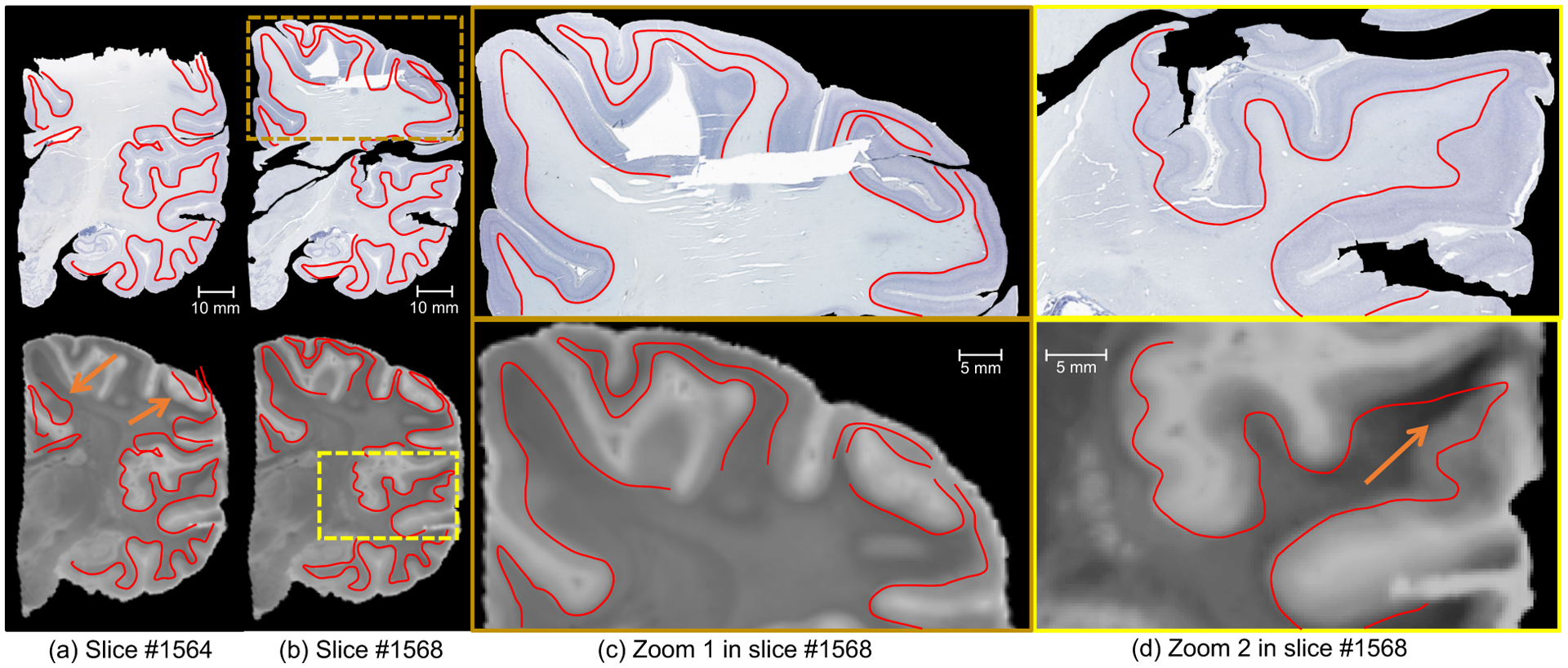
Figure 10:

Coronal view of Nissl staining and MRI for two consecutive sections (number 1564 in (a), number 1568 in (b)), in the presence of artefacts, at high-resolution (8μm). The closeup in (c) shows a well registered area from section 1568 with large registration errors in section 1564 due to severe artefacts (missing tissue), while the closeup in (d) focuses on a region with typical histology artefacts (cracks, holes). We have manually traced the white matter surface in the histology and displayed it on the registered MRI. The images show that the proposed method is not only robust but also yields accurate reconstructions. Arrows indicate large registration errors due to data artefacts, which are neither propagated to the rest of the image nor to the neighbouring sections.
