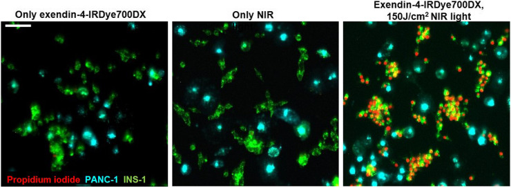
FIGURE 3.
Fluorescence microscopy of INS-1 cells labeled with fluorescent dye DiO (green) and PANC-1 cells labeled with fluorescent dye DiD (cyan), cocultured and incubated with propidium iodide (red), after incubation of exendin-4-IRDye700DX or only binding buffer and with and without NIR irradiation with radiant exposure of 150 J/cm2. Scale bar denotes 100 μm.

