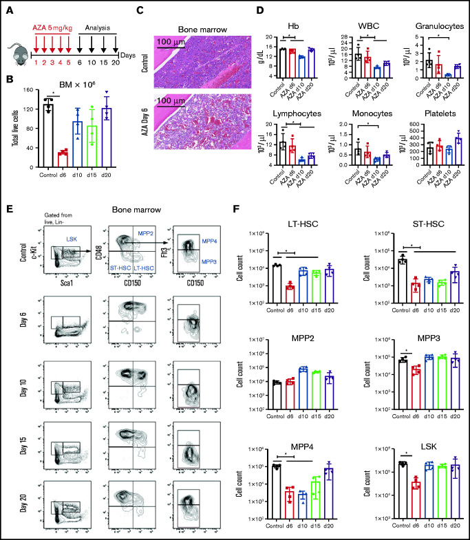
Figure 1.
AZA depletes HSPCs in vivo. (A) Treatment schema. C57BL/6 mice were injected intraperitoneally with AZA at 5 mg/kg per day for 5 consecutive days. PB, spleens, and BM from both legs and spine were analyzed on days 6, 10, 15, and 20 after the start of AZA treatment. (B) Total live cells in the BM of untreated mice and AZA-treated mice on days 6, 10, 15, and 20 after the start of AZA treatment. (C) Hematoxylin and eosin staining of a BM section from 1 mouse femur on day 6 after treatment with AZA 5 mg/kg per day for 5 days compared with an untreated control. (D) PB cell counts for untreated mice compared with AZA-treated mice on days 6, 10, and 20 after the start of AZA treatment. (E) Representative flow cytometry contour plots of the HSPC compartment in the BM of untreated and AZA-treated mice on days 6, 10, 15, and 20. The figure shows our gating strategy beginning with Lin– live cells, LSK cells, MPPs, LT-HSCs, and ST-HSCs. (F) Absolute cell counts from the different HSPC compartments on days 6, 10, 15, and 20 after AZA treatment compared with untreated control mice. LSK: Lin–Sca1+c-Kit+; LT-HSC: LSKCD150+CD48–; ST-HSC: LSKCD150–CD48–; MPP2: LSKCD150+CD48+; MPP3: LSKCD150–CD48+Flt3–; MPP4: LSKCD150–CD48+Flt3+. Data are expressed as mean ± standard deviation (SD); n = 4 mice per group per time point. *P < .05. Hb, hemoglobin; WBC, white blood cell.

