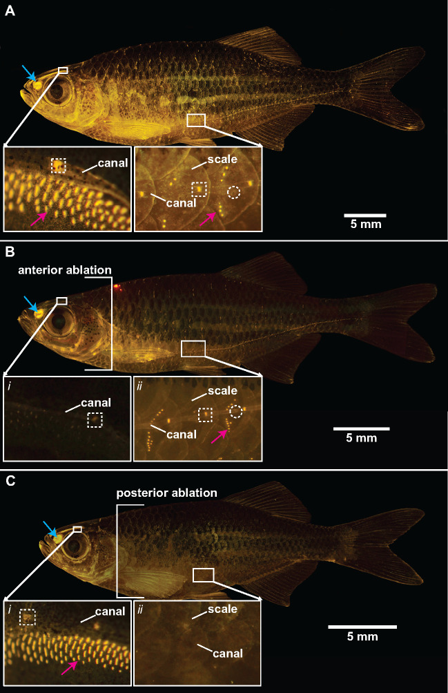
Fig. 2.
Fluorescent staining confirms lateral line ablation treatments in giant danios using cobalt chloride. Adult giant danio stained with 4-di-2-asp, showing metabolically active neuromasts of the lateral line system as bright yellow dots. (A) Before ablation, (B) anterior head ablated, and (C) posterior trunk ablated. In each panel, the inset figures show close-ups of the lateral line system in approximately the same regions on the head and trunk. Canal neuromasts shown in white dashed boxes, superficial neuromasts indicated with arrows (red), and white-dashed circles highlight canal pores. Also present are labeled olfactory cells (blue arrow). Scale bars, 5 mm.

