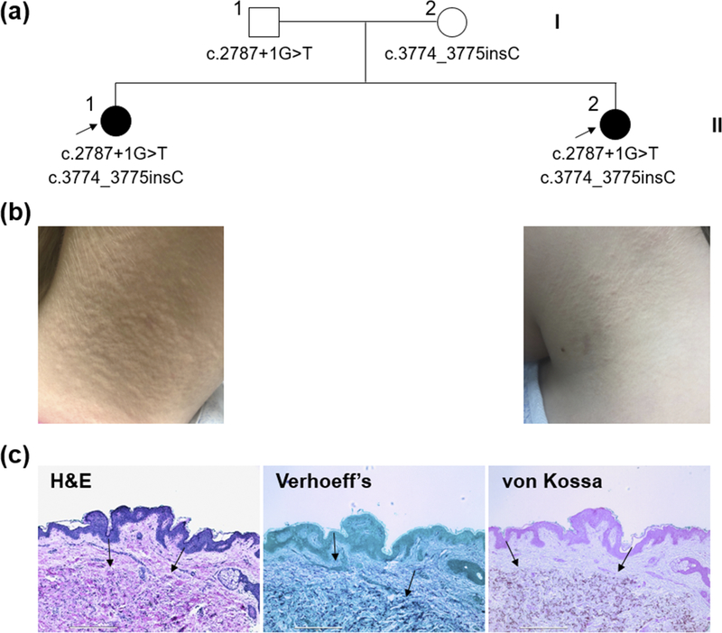
Figure 1. Nuclear pedigree and clinical features of the patients.
(a) Probands are indicated by arrows and the ABCC6 mutations are annotated under each family member. (b) Physical examination revealed yellowish papules on the bilateral neck of the older (II-1) and younger (II-2) sister, characteristic of PXE. (c) Histopathology of lesional skin in the older sister (II-1). H&E and von Kossa stains revealed mineral deposition in the mid-dermis (arrows). Verhoeff’s stain showed basophilic abnormal elastic structures in the dermis (arrows). Scale bar, 0.4 mm.

