
Figure 3.
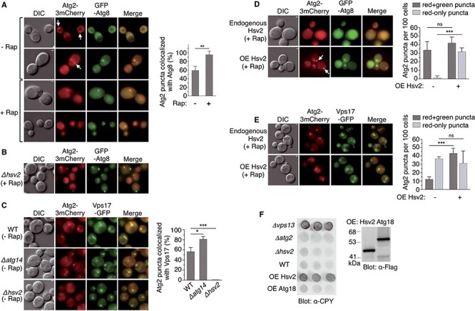
Hsv2-dependent localization of Atg2 to endosomes under basal conditions. (A, B) Colocalization of Atg2 with the PAS marker GFP-Atg8. (A) OVY384 was cotransformed with pAtg2-3mCherry and pGFP-Atg8 and examined by fluorescence and DIC microscopy in the absence or presence of rapamycin. White arrows indicate Atg2 puncta that do not colocalize with Atg8. The percentage of Atg2 puncta colocalized with Atg8 was quantified as in Fig. 1C and is shown on the right. The graph represents the mean of three experiments ± SD. **P < 0.01. (B) OVY499 (Δhsv2) expressing Atg2-3mCherry was transformed with pGFP-Atg8. Exponentially growing cells were treated with rapamycin and examined as above. (C) Colocalization of Atg2 with the endosomal marker Vps17-GFP. OVY503 (WT), OVY510 (Δatg14) and OVY543 (Δhsv2) expressing Atg2-3mCherry and Vps17-GFP were grown to mid-log phase and examined as above. The percentage of Atg2 puncta colocalized with Vps17 is shown on the right. The graph represents the mean of three experiments ± SD. *P < 0.05, ***P < 0.001. (D, E) Effect of HSV2 overexpression (OE) on Atg2 localization in rapamycin-treated cells. (D) OVY384 was cotransformed with pAtg2-3mCherry, pGFP-Atg8 and either pADH1-Hsv2(H) (OE Hsv2) or an empty vector (endogenous Hsv2). Exponentially growing cells were treated with rapamycin and examined as above. White arrows indicate Atg2 puncta that do not colocalize with Atg8. The number of Atg2-mCherry puncta that colocalize (red + green) or not (red-only) with GFP puncta was quantified as in Fig. 1C and is shown on the right. The graph represents the mean of three experiments ± SD. ***P < 0.001, ns: non-significant. (E) The same experiment was performed with OVY503 expressing Atg2-3mCherry and Vps17-GFP, transformed with either pADH1-Hsv2(U) or an empty vector. (F) Overexpression of HSV2 leads to CPY secretion. Y00000 was transformed with pADH1-Flag-Hsv2 (OE Hsv2), pADH1-Flag-Atg18 (OE Atg18) or an empty vector (WT), while OVY375 (Δvps13), OVY384 (Δatg2) and OVY380 (Δhsv2) were transformed with the empty vector. (Left) Transformants were analyzed for CPY secretion. (Right) Protein extracts from the same transformants were immunoblotted with anti-Flag Ab to detect Flag-Hsv2 and Flag-Atg18.