
Figure 6.
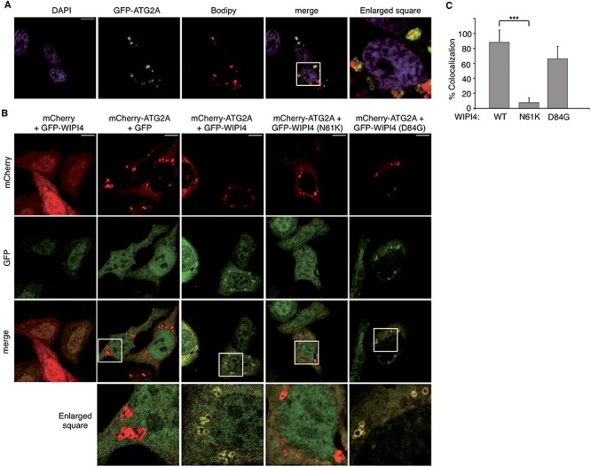
Effect of the WIPI4 substitutions N61K and D84G on the colocalization of WIPI4 and ATG2A. (A) Representative confocal fluorescence images of HeLa cells transfected with pGFP-ATG2A and stained for lipid droplets with Bodipy and nuclei with DAPI. (B) Representative confocal fluorescence images of HeLa cells cotransfected with pmCherry or pmCherry-ATG2A and pEGFP-C3, pEGFP-WIPI4 or the indicated mutant derivatives. GFP, mCherry and DAPI fluorescence channels are shown. Scale bar 10 μm. White box represent enlarged area shown. (C) Percentage of mCherry-ATG2A structures that colocalize with GFP-WIPI4. Data represent means ± SD of at least 283 mCherry-ATG2A positive structures in 87 co-transfected cells from five independent transfections. P values were determined by unpaired two-tailed Student’s t-test. ***P < 0.001.