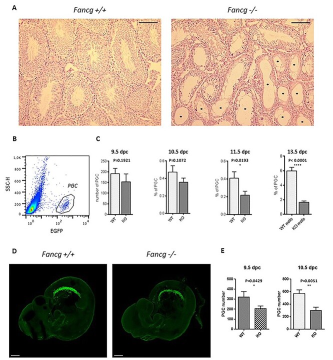
Figure 1 .

Fancg −/− mice exhibit loss of germinal cells in embryos and adult testis. (A) Hematoxylin–eosin-stained histological sections of testes of adult 3-month-old Fancg+/+ and Fancg−/− mice. Fancg−/− testis showed germinal aplasia with SCO tubules (*) (scale = 100 μm). (B) EGFP-positive PGC identification in E13.5 OG2:Fancg+/+ embryos using flow cytometry. (C) Quantification of the absolute number of PGCs per embryo using Trucount microbeads at E9.5 dpc wild-type (WT) n = 21, knockout (KO) n = 10), of the frequency of PGCs per embryo at E10.5 (WT n = 17, KO n = 15), and of the frequency of PGCs per gonads at E11.5 (WT n = 12, KO n = 11) and E13.5 (WT n = 8, KO n = 9). (D) Confocal microscopy analysis of E10.5 cleared embryos using an antibody against EGFP to identify PGCs (scale = 300 μm). (E) Automated cell counting of PGCs per embryo using Imaris software at E9.5 (WT n = 4, KO n = 5) and E10.5 (WT n = 4, KO n = 5).
