Figure 5 .

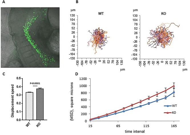
OG2:Fancg −/− PGCs also exhibited increased cell speed and mean square displacement (MSD) in ex vivo E9.5 embryo culture. (A) Ex vivo migration assay of PGCs. E9.5 embryos were cultured and filmed for 8–12 h, EGFP fluorescence and diffusion light frames were captured every 15 min. (B) Movement of OG2:Fancg−/− and OG2:Fancg+/+ PGCs shown from the same origin point. (C) Average cell speed and (D) MSD of tracked PGCs from ex vivo culture of E9.5 OG2:Fancg+/+ and OG2:Fancg−/− embryos (P < 0.005).
