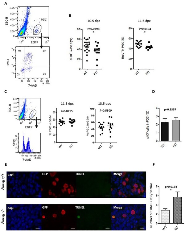
Figure 2 .

Decreased proliferation rate and increased cell death of PGCs in Fancg−/− embryos at E11.5. (A) Identification of the proliferative BrdU-positive cell fraction (Q1 and Q2) in the EGFP-positive PGC population from E11.5 WT embryos. (B) Quantification of the proliferative fraction of PGCs at E10.5 (WT n = 17, KO = 11) and E11.5 (WT n = 14, KO = 15) in OG2:Fancg+/+ and OG2:Fancg−/− embryos. (C) DNA content analysis (7-AAD) of the EGFP-positive PGC population at E11.5 (WT n = 5, KO n = 4) and E13.5 (WT n = 11, KO n = 10). (D) Frequency of mitotic PHH3-positive PGCs counted from histological sections of testes in E13.5 OG2:Fancg+/+ and OG2:Fancg−/− embryos (WT n = 3, KO n = 3). (E) Detection of cell death by TUNEL assays in histological sections of E11.5 OG2:Fancg+/+ and OG2:Fancg−/− embryos. Nuclei counterstained by DAPI (blue), PGCs labeled with an antibody against EGFP (red) and the TUNEL-positive signal (green) are shown (scale = 10 μm). (F) Quantification of the number of TUNEL-positive dying cells per section at E11.5 (WT n = 6, KO n = 6).
