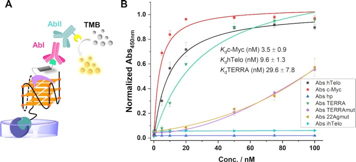
Figure 4.
(A) Schematic representation of 5-BrdU PDC conjugate recognition by antibodies and chromogenic detection in vitro (ELISA, TMB = 3,3′,5,5′-Tetramethylbenzidine). (B) Binding curves determined by an adapted ELISA assay for hTelo, c-Myc, and TERRA as examples of G4s, a hairpin (hp) as example of a duplex, 22Agmut and TERRAmut as examples of G-rich sequences not able to fold in a G4 structure, and ihTelo as example of a C-rich sequence (normalized Abs450nm of TMB versus oligonucleotide concentration) and dissociation constants (Kd) obtained from curve fitting (1:1) in the presence of G4 sequences. Error bars represent the SD calculated from three replicates.

