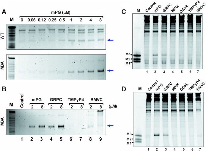
Figure 7.

Effects of ligands on the stabilization of PDGFR-β GVBQ in the G-rich ssDNA and dsDNA. (A) Protection of WT and M3A ssDNA from being hydrolyzed by T4 DNA polymerase in the presence of mPG. The blue arrow indicates the cleavage band before GVBQ as shown in Figure 2B. (B) Compare the protection effects of mPG, GRPC, TMPyP4 and BMVC on M3A ssDNA from being hydrolyzed by T4 DNA polymerase. The blue arrow indicates the cleavage band before GVBQ as in Figure 2B. (C) Detecting the effects of mPG and other compounds (2 μM) on the stability of PDGFR-β GVBQ in transcribed WT plasmids by RNA polymerase arrest. (D) Detecting the effect of mPG and other compounds (2 μM) on the stability of PDGFR-β GVBQ in transcribed M3A plasmids by RNA polymerase arrest. Marker (M) shows termination sites as labeled in Figure 3.
