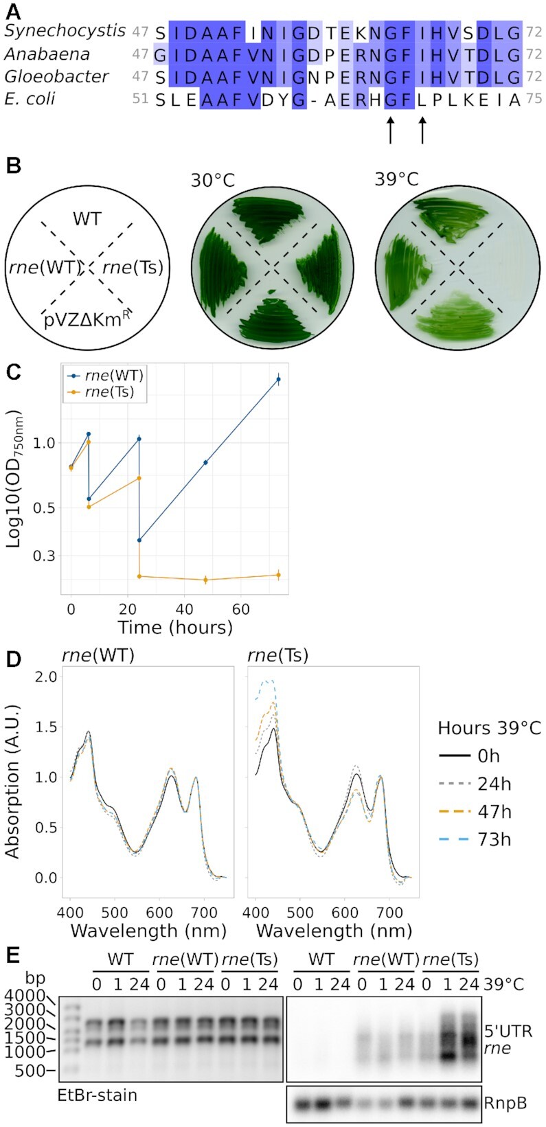
Figure 1.

Characterisation of Synechocystis harbouring temperature-sensitive RNase E. (A) Alignment of Synechocystis RNase E residues 47–72 with the respective section in homologues from Anabaena sp. PCC 7120, Gloeobacter violaceus PCC 7421 and E. coli. The arrows point at two conserved residues which were mutated to obtain temperature sensitivity. (B) Growth of wild-type Synechocystis (WT), rne(WT), rne(Ts) and an empty-vector control strain (pVZΔKmR) at the standard temperature of 30°C and at 39°C. The scheme on the left indicates the streaking order of the strains. (C) Growth of rne(WT) and rne(Ts) at 39°C in liquid culture. Time point 0 h corresponds to the switch from 30°C to 39°C. Error bars indicate the standard deviation of biologically independent duplicates. Cultures were diluted after 6 hours and 24 hours of growth. (D) Absorption spectra of rne(WT) and rne(Ts) throughout the course of 73 h incubation at 39°C of one representative experiment (compare panel C). Spectra were normalized to absorption at 682 nm and 750 nm. (E) Northern blot analysis of the accumulation of the rne-rnhB transcript. One representative analysis is shown (n = 4). In addition to the control hybridization with RnpB (lower panel), the denaturing agarose gel which was used for blotting is shown on the left as a loading control.
