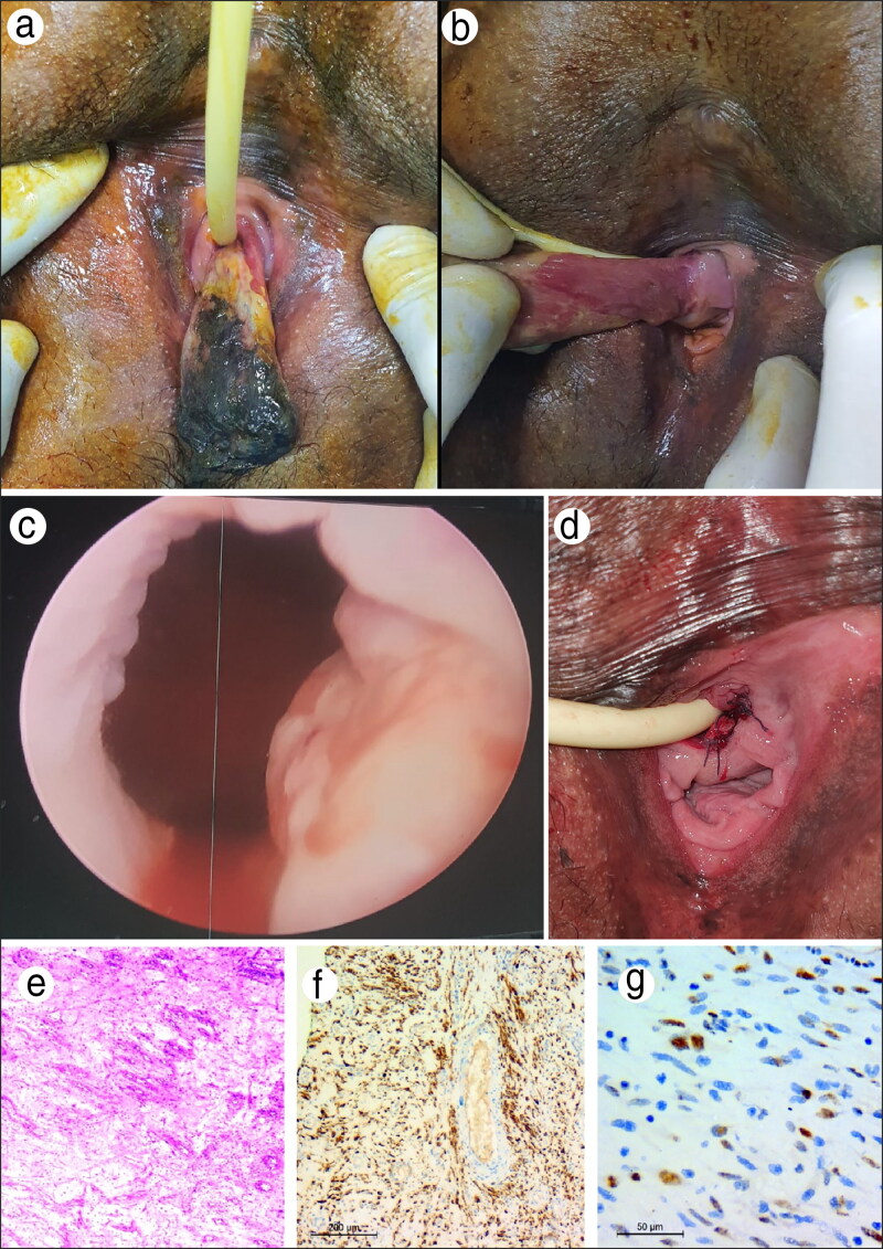
Figure 1.
(a, b) The relation of the mass to the urethra with urethral catheter. (c) Cystoscopy showing suspicious mucosa at the bladder neck. (d) Postoperative picture after the mass was excised. (e–g) Histopathology showing (e) a hypocellular lesion with bland-looking neoplastic spindle to stellate cells with small-sized blood vessels in a myxoid background, with neoplastic cells showing immunoreactivity for (f) desmin and (g) estrogen receptor.

