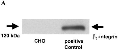
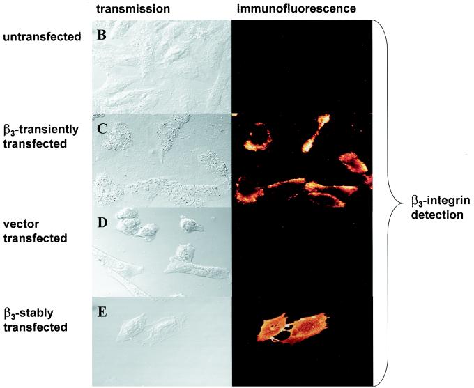
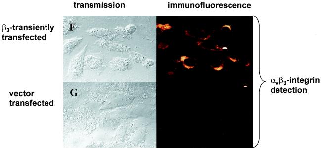
FIG. 1.
Detection of β3-integrin in CHO cells. (A) Western blot. Cell lysates were resolved on a 10% polyacrylamide gel, and β3-integrin was detected by Western blot analysis using an antibody to β3- integrin. The band at ∼120 kDa represents β3-integrin. The β3-integrin-expressing ovarian cancer cell line OVMZ-6 served as a positive control. The experiment was repeated twice. (B to G) Immunofluorescence. Untransfected CHO cells (B), CHO cells transiently transfected with β3-integrin (C and F) or the β3-integrin vector (D and G), or a stable β3-integrin-expressing CHO cell clone, A5 (E), was plated on chamber slides and stained by indirect immunofluorescence. CHO cells were incubated with either a monoclonal antibody against β3- integrin (B to E) or an antibody against αvβ3-integrin (F and G) followed by an Alexa 488-conjugated goat anti-mouse secondary antibody. The experiment was repeated twice.


