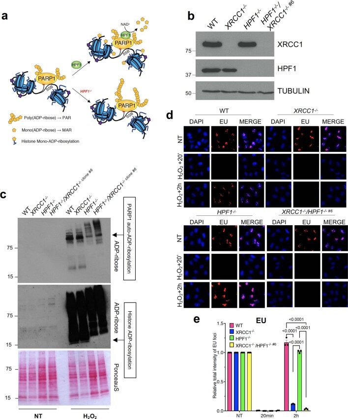
Extended Data Fig. 7. Deletion of the histone ADP-ribosylation factor HPF1 does not rescue transcription recovery in XRCC1−/− RPE-1 cells.
a, Model of the impact of HPF1 on PARP1-mediated histone ADP-ribosylation as well as PARP1 auto-ADP-ribosylation. b, Immunoblot of HPF1 and XRCC1 in WT, HPF1−/−, XRCC1−/−, and HPF1−/−/XRCC1−/− U2OS cells. A representative blot from one of three independent experiments is shown. c, Immunoblot of ADP-ribosylation upon H2O2 treatment (1 mM, 5 min) in WT, HPF1−/−, XRCC1−/−, and HPF1−/−/XRCC1−/− U2OS cells. A representative blot from one of three independent experiments is shown. d, Global transcription (EU Immunofluorescence) in WT, HPF1−/−, XRCC1−/−, HPF1−/−/XRCC1−/− U2OS cells following mock treatment or at the indicated times after treatment with 1 mM H2O2 for 20 min. Scale bars, 10 μm. e, Quantification of the EU signal shown in (d). Data are means (±s.e.m.) of three independent experiments, and statistically significant differences were determined by two-way ANOVA with Tukey’s multiple comparisons test (p values are indicated).

