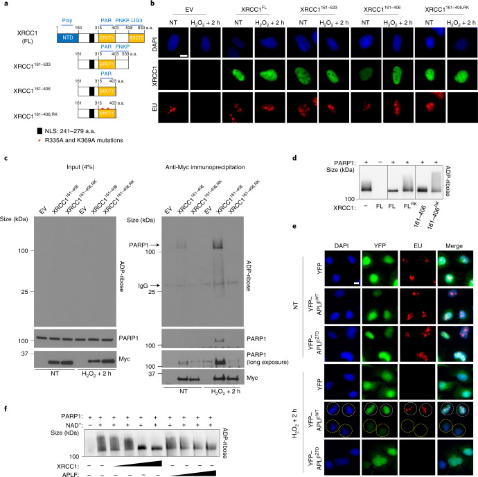
Fig. 4. The poly(ADP-ribose)-binding activity of XRCC1 promotes transcriptional recovery following oxidative damage.
a, Cartoon of full-length (FL) and truncated XRCC1 proteins encoded by the Myc- and/or His-tagged expression constructs employed in this work. The interaction partners are shown (top): Polβ, DNA polymerase β; PAR, poly(ADP-ribose); PNKP, polynucleotide kinase phosphatase; LIG3, DNA ligase III. The N-terminal domain (NTD), nuclear localization signal (NLS) and two BRCT domains are also shown; a.a., amino acids. b, Representative images showing the levels of global transcription (EU pulse labelling; bottom) in XRCC1−/− U2OS cells, transiently transfected with expression constructs encoding the indicated XRCC1 proteins, following mock treatment or 2 h after treatment with 1 mM H2O2 for 20 min. The XRCC1 protein levels are also shown (middle). The cells were pulse labelled with EU as in Fig. 1. EV, empty vector. c, Immunoblot of Myc-tagged XRCC1 proteins, PARP1 and ADP-ribose levels in cell extract from the indicated transiently transfected XRCC1−/− U2OS cells before (left, input) and after (right) anti-Myc immunoprecipitation, 2 h after mock treatment or treatment with 1 mM H2O2 for 20 min (right). d, XRCC1 suppresses PARP1 auto-ADP-ribosylation in vitro. Human recombinant PARP1 (100 nM) was incubated in the presence of single-stranded DNA (100 nM), NAD+(2.5 μM) and with or without 1 μM of full-length XRCC1 or the indicated XRCC1 protein fragment for 30 min. The reaction products were fractionated by SDS–PAGE and detected by western blotting with ADP-ribose detection reagent. e, Representative images showing the levels of global transcription (EU pulse labelling) in XRCC1−/− U2OS cells stably transfected with expression constructs encoding WT APLF (yellow fluorescent protein (YFP)–APLFWT) or its ADP-ribose binding mutant (YFP–APLFZFD) following mock treatment or 2 h after treatment with 1 mM H2O2 for 20 min. White and yellow circles indicate examples of cells over-expressing or not YFP-APLF, respectively. For b,e, representative images from one of three independent experiments are shown. Scale bars, 10 μm. f, APLF suppresses PARP1 auto-ADP-ribosylation in vitro. Human recombinant PARP1 (100 nM) was incubated in the presence of single-stranded DNA (100 nM), NAD+(2.5 μM) and with or without 0.25, 0.5, 1 or 1.5 μM WT recombinant human APLF or XRCC1 for 30 min. The reaction products were fractionated by SDS–PAGE and detected by western blotting with ADP-ribose detection reagent. c,d,f, Representative blots from one of three independent experiments are shown.

