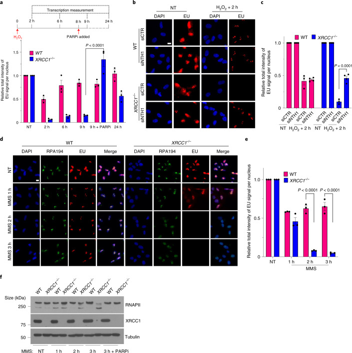
Fig. 5. Unrepaired BER intermediates are a source of toxic PARP1 activity and transcriptional suppression in XRCC1-defective cells.
a, Levels of global transcription (EU immunofluorescence; bottom) from the experiment outlined in the schematic (top; see also Extended Data Fig. 6a). b, Representative images of the levels of global transcription (EU pulse labelling) in WT and XRCC1−/− RPE-1 cells following mock treatment or at 2 h after treatment with 250 μM H2O2 for 5 min. The cells were pretreated with control (siCTR) or siRNA targeting NTH1 (siNTH1) for 72 h. The cells were pulse labelled with EU as in Fig. 1. c, Levels of global transcription from the experiment in b. d, Representative images of the levels of global transcription (EU pulse labelling) in WT and XRCC1−/− RPE-1 cells following mock treatment or at the indicated times after treatment with 0.1 mg ml−1 MMS (continuous treatment). The cells were pulse labelled with EU as in Fig. 1. b,d, Scale bars, 10 μm. e, Levels of global transcription (EU pulse labelling) from the experiment shown in d. a,c,e, Data are the mean ± s.e.m. of three independent experiments. Statistical significance was determined using a two-way ANOVA with Sidak’s multiple comparisons test (significantly different P values are indicated). f, Immunoblot of RNAPII hyperphosphorylation in WT and XRCC1−/− RPE-1 cells following mock treatment or after treatment for the indicated times with 0.1 mg ml−1 MMS. Where indicated, 10 μM PARPi was present throughout the MMS treatment. A representative blot from one of three independent experiments is shown.

