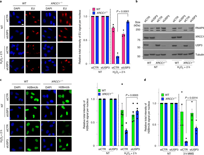
Fig. 7. Toxic PARP1 activity disrupts histone monoubiquitination and promotes transcriptional suppression via USP3.
a, Representative images (left) and levels (right) of global transcription (EU pulse labelling) in WT and XRCC1−/− RPE-1 cells following mock treatment or at the indicated times after H2O2 treatment (250 μM; 5 min). The cells were pretreated with control (siCTR) or USP3 siRNA (siUSP3) and pulse labelled with EU. b, RNAPII hyperphosphorylation in WT and XRCC1−/− RPE-1 cells, pretreated with control or USP3 siRNA as indicated, following mock treatment or 2 h after treatment with H2O2 as in a. A representative blot of the whole-cell extracts from one of three independent experiments is shown. c, Representative images (left) and levels (right) of H2BmUb in WT and XRCC1−/− RPE-1 cells, pretreated with control or USP3 siRNA as in a following H2O2 treatment. a,c, Scale bars, 10 μm. d, Levels of H2BmUb in WT and XRCC1−/− RPE-1 cells following mock treatment or treatment with 0.1 mg ml−1 MMS for 3 h. The cells were pretreated with control or USP3 siRNA. a,c,d, Data are the mean ± s.e.m. of three (a), five (c) and four (d) independent experiments. Statistical significance was determined using a two-way ANOVA with Tukey’s multiple comparisons test (significantly different P values are indicated).

