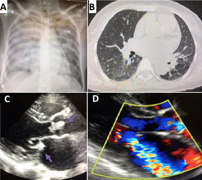
Figure 2.
clinical case 2; A) chest X-ray showed a bilateral diffuse alveolar syndrome and cardiomegaly; B) thoracic CT scan a cardiomegaly with the presence of peripheral, central and bilateral ground glass lesion; C) transthoracic echocardiogram: parasternal long axe view showing a vegetation in the aortic and mitral valve (arrows); D) Doppler signal showing mitral regurgitation

