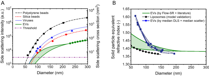
Figure 1.
(A) Side scattering intensity measured (symbols) by a CytoFLEX1 and calculated (lines) by Mie theory10 versus diameter for polystyrene beads (squares), silica beads (circles), viruses (triangles), and CD61 + EVs (hexagons). The assumed refractive index (RI) is 1.627 for polystyrene beads, 1.440 for silica beads, and 1.472 for viruses at the illumination wavelength of 405 nm. EVs were modelled as core–shell particles, having a 12 nm thick shell with an RI of 1.52 and a core RI of 1.353 (upper solid line), and having a 4 nm thick shell with an RI of 1.450 and a core RI of 1.380 (lower solid line). The horizontal line indicates the trigger threshold. (B) Solid particle equivalent RI versus diameter for plasma-derived EVs based on Flow-SR11 and literature (panel A, solid green lines), for liposomes19 (dotted line), and for plasma-derived EVs based on the dynamic light scattering (DLS) measurements and the flow cytometry light scattering measurements performed by Brittain et al.1 (overlapping blue lines and symbols). Below a diameter of 100 nm, the solid particle equivalent RI based on DLS and flow cytometry light scattering is substantially higher compared to estimates based on Flow-SR11 and literature and liposome measurements.

