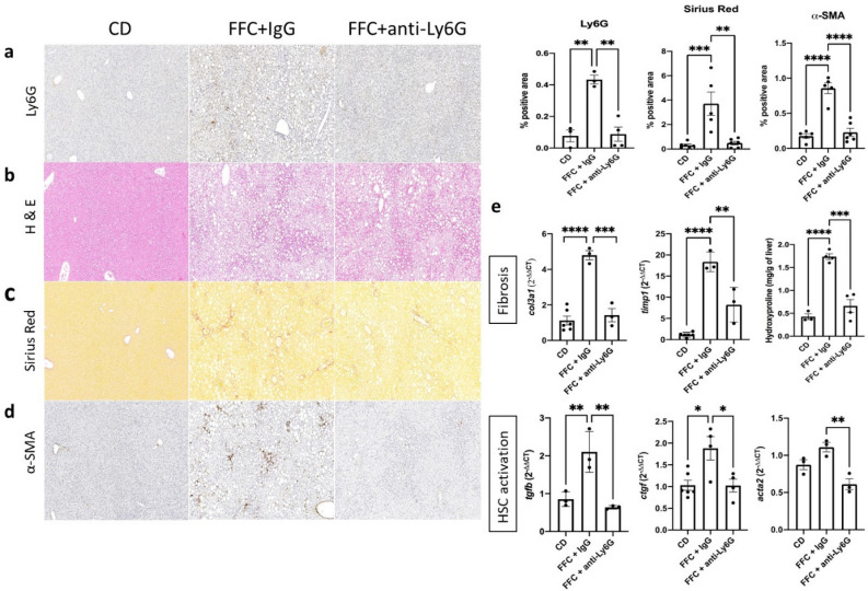
Figure 2.
Hepatic steatosis, fibrosis and hepatic stellate cell (HSC) activation are ameliorated in neutrophil depleted mice during NASH development. Neutrophil marker Ly6G (a), steatosis (b) and fibrosis (c) levels were assessed via IHC, H&E and Picrosirius Red stainings IHC respectively on paraffin-embedded tissue sections. Activated α-SMA+ HSCs (d) were detected via IHC. Quantification and ANOVA comparison were made between the control diet and IgG or anti-Ly6G regimen receiving groups during ongoing NASH development. (e) mRNA expression of hepatic fibrosis and HSC activation related genes and hydroxyproline levels were analyzed between the mentioned groups and compared with ANOVA IHC. CD control diet, α-SMA/acta2 alpha smooth muscle actin, IHC immunohistochemistry, col3a1 collagen type 3 alpha 1 chain, timp1 tissue inhibitor of metalloproteinases 1, tgfb transforming growth factor beta, ctgf connective tissue growth factor.

