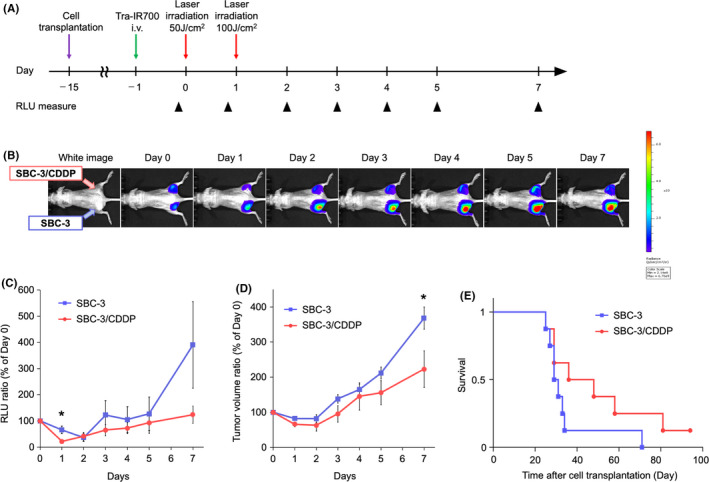
FIGURE 4.

Comparing in vivo NIR‐PIT anti‐tumor effect in SBC‐3 and SBC‐3/CDDP tumors. (A) The treatment regimen and imaging schedule. Bioluminescence images (BLI) were taken as indicated. (B) BLI of SBC‐3 (left‐side dorsum) and SBC‐3/CDDP (right‐side dorsum) tumor‐bearing mice along with the treatment. Both groups were about the same size and exhibited similar bioluminescence intensity before NIR‐PIT treatment. (C) Quantitative luciferase activity (RLU value at day 0 is set to 100%) showed a decrease in both groups after NIR‐PIT treatment. RLU ratio was lower in SBC‐3/CDDP tumors than in SBC‐3 tumors (data are means ± SEM. n = 8, *p = 0.022 < 0.05, at day 1, Student's t‐test). (D) Tumor growth (estimated tumor volume at day 0 is set to 100%) was significantly inhibited in SBC‐3/CDDP tumors compared with SBC‐3 tumors (data are means ± SEM. n = 8, *p = 0.043 < 0.05, at day 7, Student's t‐test). (E) Kaplan–Meier analysis. SBC‐3/CDDP tumor group tended to have longer survival compared with the SBC‐3 tumor group, but no statistically significant difference was detected (n = 8, p = 0.124, log‐rank test). CDDP, cisplatin; NIR‐PIT, near‐infrared photoimmunotherapy; RLU, relative light units
