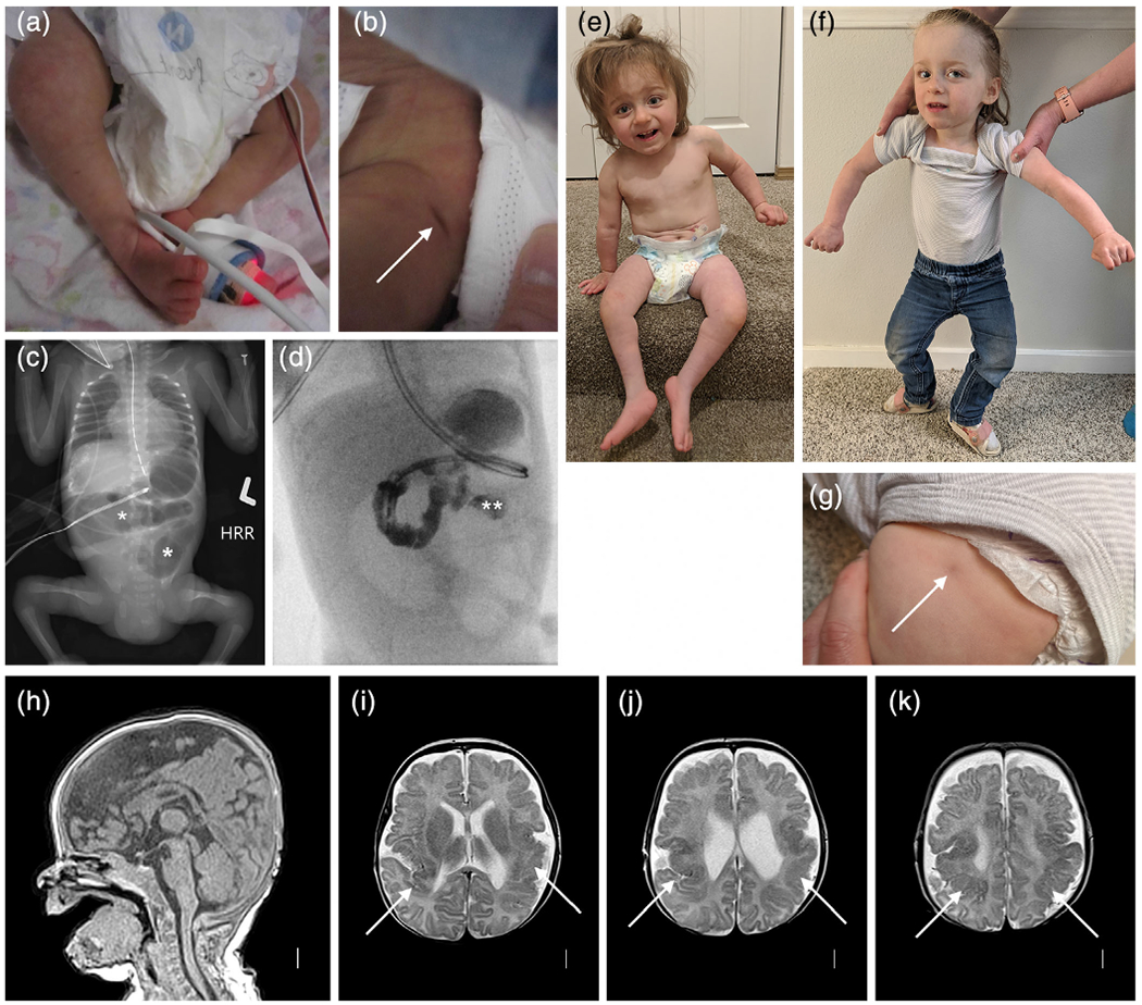
FIGURE 5.

Clinical photographs, abdominal radiography, and brain imaging from a monozygotic (MZ) twin girl (LR18-005). Photos taken in the first few days of life show right clubfoot and a dimple over her left hip (arrow in (b)). An abdominal radiograph shows abnormal loops of distended bowel distally (asterisks in (c)), concerning for obstruction. On fluoroscopic upper GI series, contrast does not pass beyond the duodenojejunal junction (double asterisk in (d)), and subsequent exploratory laparotomy confirmed jejunal atresia. Photographs at 2 and 2.5 years show small legs and bilateral clubfeet that have improved with therapy (e), persisting abnormal posture from her leg contractures as well as mild hand contractures (f), and another photo of a dimple over her left hip (g). Brain MRI (T1 sagittal and T2 axial images) show normal midline structures (h), and irregular and infolded gyral pattern consistent with polymicrogyria (PMG) in bilateral posterior frontal, perisylvian and parietal regions (arrows in (i–k)) that appears slightly more severe on the left (i.e., right side of the image) [Color figure can be viewed at wileyonlinelibrary.com]
