
FIGURE 8.
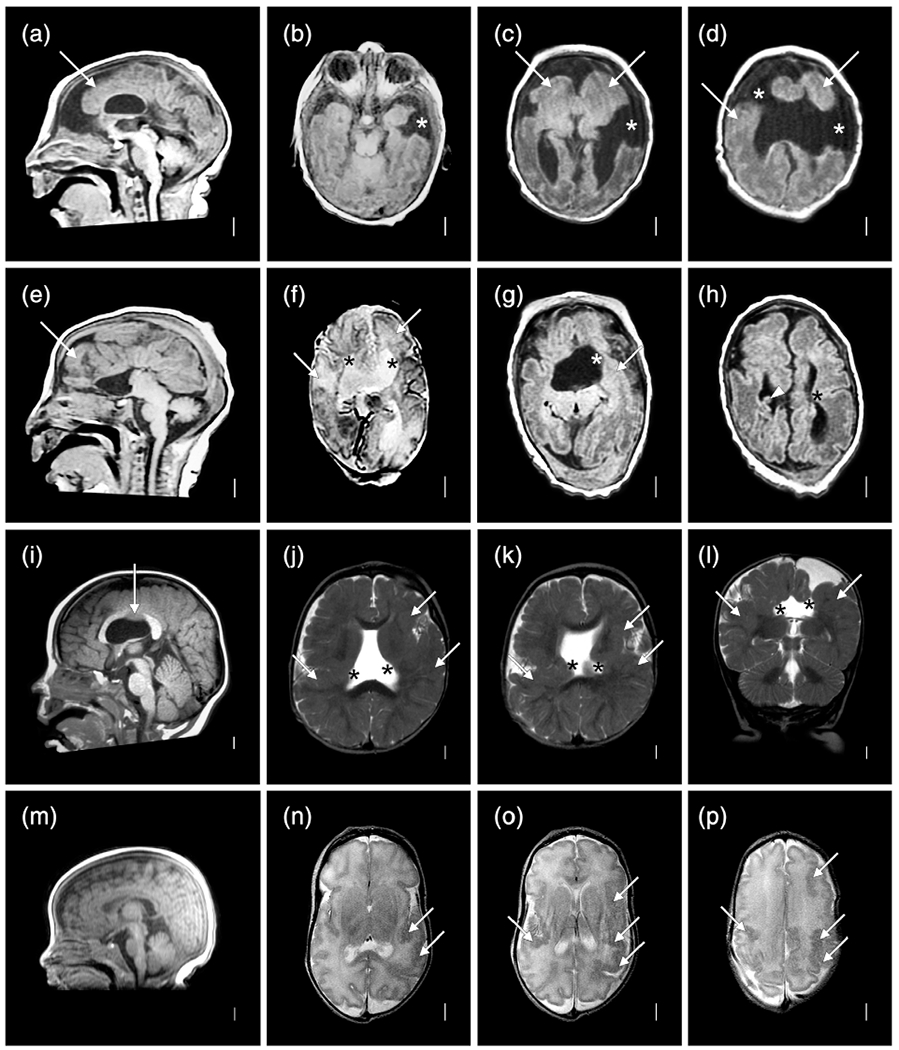
Brain MRI selected to show more diverse patterns of malformations of cortical development (MCD) despite low resolution studies in individuals LR02-073a1 (a–d), LR02-073a2 (e–h), LR01-231a1 (i–l), and LR17-510 (m–p). The co-twins in the top two rows both have microcephaly with low, sloping foreheads, extensive PMG (white arrows in (a–h)) and clefts, but the clefts are widely open in a1 (asterisks in (b–d)) and closed in a2 (asterisks in (f–h)). A prominent periventricular nodular heterotopia (PNH) is seen in a2 as well (arrowhead in (h)). The next child has extensive PMG (arrows in (j–l)) surrounding closed lip clefts (asterisks in (j–l)) associated with a missing middle segment in the body of the corpus callosum (arrow in (i)). The last child has highly asymmetric PMG more severe on the left (arrow in (n–p)) without a cleft