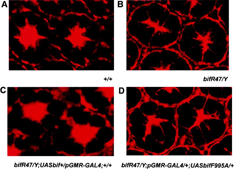
FIG. 7.
Rescue of the abnormal F-actin localization pattern of bif pupal eye discs. (A to D) Each panel shows two ommatidia from a 55-h pupal eye disc stained with tetramethyl rhodamine isothiocyanate-labeled phalloidin. (A) A wt F-actin staining pattern reveals the organization of rhabdomeres in the center of the eye. (B) A homozygous bifR47 mutant shows disorganized F-actin staining and merged rhabdomeres in the center of the eye, which is seen at a frequency of 92% in the 100 ommatidia scored in pupae. (C) Rescue of the bif pupal eye disc phenotype by pGMR-driven expression of a bif+ transgene. The expression of the wt UAS-bif transgene caused the defective F-actin staining to revert to near-wt patterns, with only 16% of the ommatidia retaining the mutant phenotype (n = 100). (D) Expression of the bifF995A mediated by the pGMR-GAL4 driver fails to rescue the defective F-actin distribution pattern associated with bifR47 allele, as 86% of the ommatidia scored retain the mutant phenotype (n = 100).

