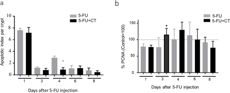
Fig. 3.
Effects of CT on the induction of apoptosis and proliferation of small intestinal crypt cells in 5-FU-induced small intestine mucositis model mice. a Apoptosis was evaluated by the TUNEL assay. TUNEL-positive cells were counted in 10 small intestinal crypts per specimen under a light microscope. Values are expressed as the mean ± SEM. N = 5–6. * p < 0.05 vs 5-FU by the two-tailed Student’s t-test. b Cell proliferation activity was immunohistochemically measured using the anti-PCNA antibody. PCNA-positive cells were counted in intestinal crypts under a light microscope. The average number of PCNA-positive cells in each individual mouse was divided by the average number of PCNA-positive cells in normal mice and expressed as % PCNA. Values are expressed as the mean ± SEM. N = 5–6. * p < 0.05 vs 5-FU by the two-tailed Student’s t-test

