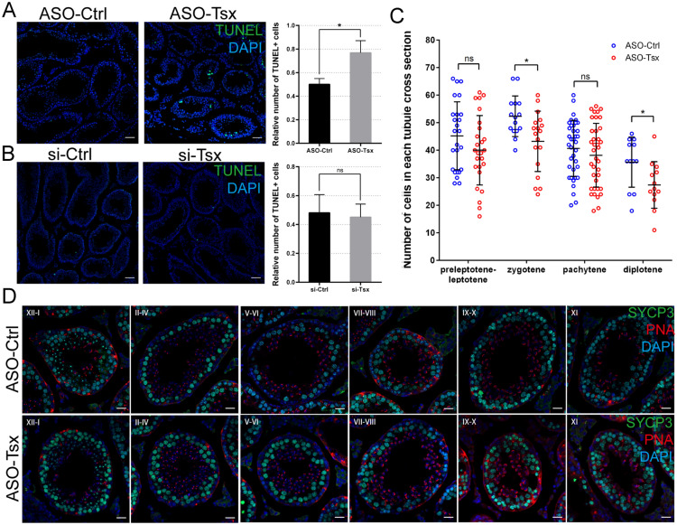
Fig. 4.
Physiological outcomes of Tsx ASOs in testis. A, B TUNEL staining and quantification of TUNEL+ cells in tubules from Tsx-knockdown mice at age of 3 weeks injected with ASOs (A) or siRNAs (B). Nuclei were stained with DAPI. Scale bar, 50 μm. Relative number of TUNEL+ cells is determined by counting the number of TUNEL+ cells per field and dividing by the number of tubules in each field. 37 fields including 150 tubules from ASOs-mediated Tsx-knockdown testes, 31 fields including 125 tubules from ASO-control testes, 21 fields including 100 tubules from siRNAs-mediated Tsx-knockdown testes and 30 fields including 111 tubules from siRNA-control were counted. Each contains three independent samples. *p < 0.05. C Quantification of four cell populations between control and Tsx-knockdown testes. Number of each cell population per tubule is shown in dot. 81 tubules from 3 testes injected with ASO-Tsx and 83 tubules from 3 testes injected with control ASO were analyzed. *p < 0.05. D Immunostaining of SYCP3 (green) and PNA (red) on testis sections from control and Tsx-knockdown mice. Seminiferous tubule sections were staged by morphology of chromosome and acrosome. Scale bar, 20 μm. A–C Values are expressed as mean ± SD. Statistical significance was determined using t tests

