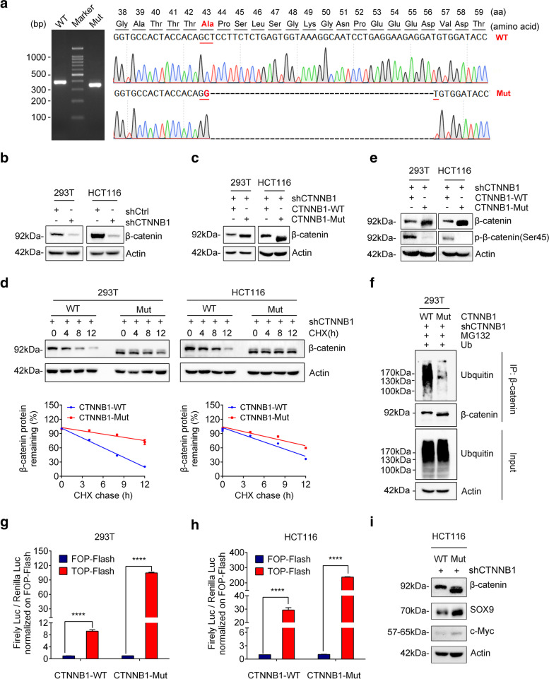
Fig. 4.
CTNNB1-Mut promotes Wnt/β-catenin signaling pathway via increasing the stability of β-catenin. (a) Agarose gel electrophoresis of PCR products generated by the cDNA of CP111 tissue (left panel) and schematic representation of CTNNB1 deletion mutants generated from ACP patient cells. WT: CTNNB1 wild type; Mut: CTNNB1 mutation type. (b) Knockdown efficiency of CTNNB1 shRNA (shCTNNB1) in both 293 T and HCT116 cells analyzed by western blotting. (c) Western blotting was performed to verify the expression of CTNNB1-WT/Mut 293 T/HCT116 cells. (d) CTNNB1-WT/Mut 293 T/HCT116 cells were cultured in the presence of CHX (50 μg/ml) for 0, 4, 8, 12 h, followed by immunoblotting (IB) using anti-β-catenin and actin antibodies. (e) CTNNB1-WT/Mut 293 T/HCT116 cells were treated with 50 nM Calyculin A for 30 min. The cell lysate was IB with anti-β-catenin and anti-β-catenin-phosphate-S45 antibodies. (f) Ubiquitin was transfected into 293 T cells which respectively infected with CTNNB1-WT or CTNNB1-Mut. After 48 h, cells were treated with 10 μM MG132 for 10 h. Cell lysates were subjected to IP with anti-β-catenin followed by IB with ubiquitin or anti-β-catenin antibody. (g-h) TOP-Flash reporter or FOP-Flash reporter was co-transfected with pRL-TK plasmids into CTNNB1-WT/Mut 293 T/HCT116 cells. Luciferase activity was measured with the Dual-Luciferase reporter assay, and relative luciferase activity was normalized to the FOP-Flash of the CTNNB1-WT group. Values are mean ± SD for triplicate samples. (i) Levels of SOX9 and c-Myc protein in HCT116-shCTNNB1 cells infected with CTNNB1-WT or CTNNB1-Mut were determined by Western blotting

