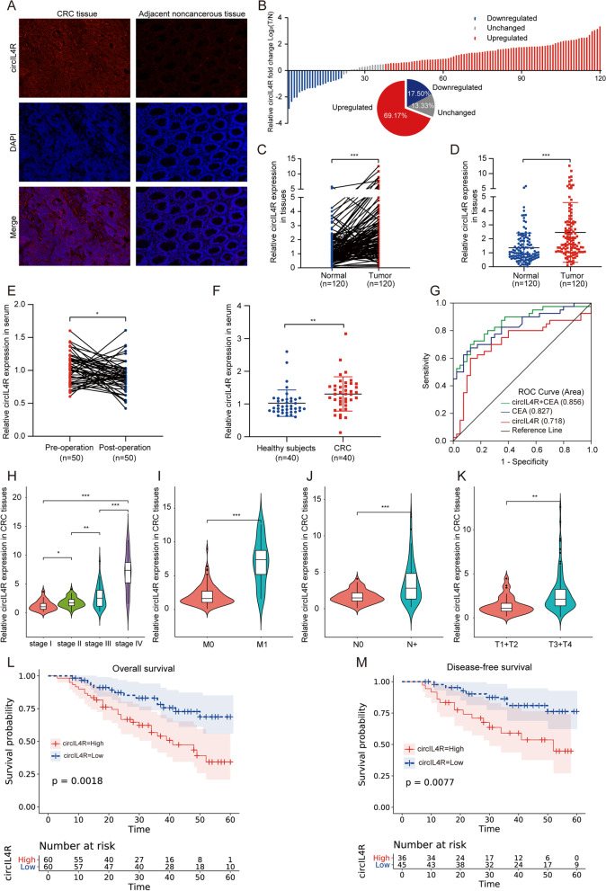
Fig. 2.
Clinical significance of circIL4R in CRC tissues and serum. a FISH staining assay showed that circIL4R expression in CRC tissues was higher than that in the corresponding ANTs (200×). b-d qRT-PCR analysis showed that circIL4R expression was significantly upregulated in 120 CRC tissues compared with paired ANTs. e Changes in the levels of serum circIL4R in 50 newly diagnosed CRC patients before and after surgery were detected by qRT-PCR. f Relative levels of serum circIL4R in healthy controls (n = 40) and CRC patients (n = 40) were determined by qRT-PCR. g ROC curves were used to determine the diagnostic value of serum circIL4R either alone or in combination with CEA in CRC. h-k circIL4R expression was evaluated in different groups stratified according to clinical characteristics (pathological stage, M classification, N classification, T classification.) by violin plot. l and m Kaplan–Meier survival curves showed that CRC patients with high circIL4R expression exhibited shorter OS (n = 120, P = 0.0018) and DFS (n = 120, P = 0.0077) based on 120 clinical specimens. *P < 0.05, **P < 0.01, ***P < 0.001

