Fig. 9.
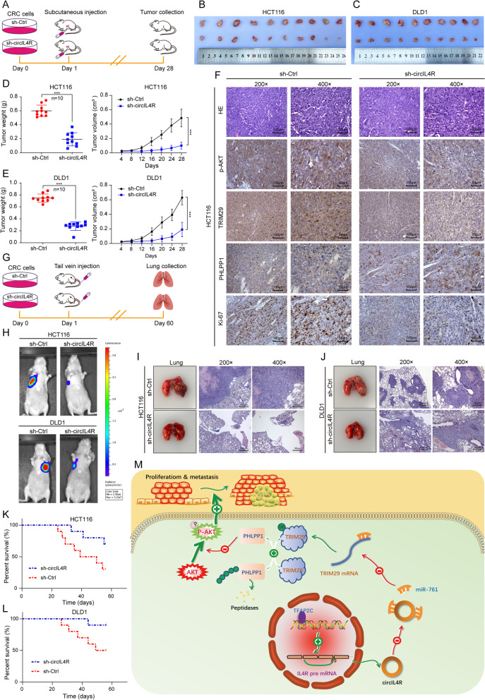
circIL4R promotes the tumorigenesis and metastasis of CRC cells in vivo. a Schematic illustration of the subcutaneous xenograft tumor model in BALB/c nude mice. b and c Images of dissected subcutaneous xenograft tumors from different groups. d and e Growth curve and weight analysis of xenograft tumors in nude mice. f Sections of xenograft tumors were subjected to H&E and IHC staining using Ki67, TRIM29, PHLPP1, and p-AKT antibodies. g. Schematic illustration of the tail vein-injected nude mouse model. h Representative images and luminescence intensity of lung metastatic nodules in tail vein-injected nude mice. (n = 5 per group). i and j Representative images and HE staining of dissected lungs and metastatic nodules. k and l The survival curves of nude mice injected with CRC cells were determined by the Kaplan–Meier method. m Schematic illustration of the mechanism by which circIL4R activates the PI3K/AKT signaling pathway via the miR-761/TRIM29/PHLPP1 axis and promotes proliferation and metastasis in CRC. *P < 0.05, **P < 0.01, ***P < 0.001

