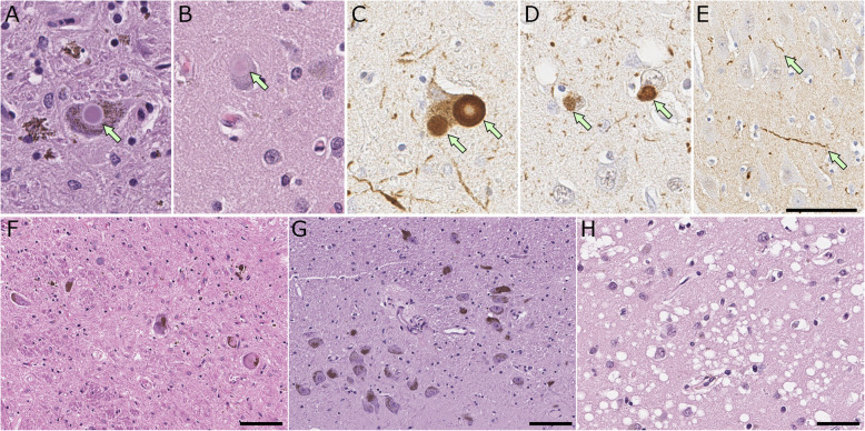
Fig. 4.
Representative images of histopathology of Lewy body disease. A, B, F-H hematoxylin and eosin staining, C-E immunohistochemistry for α-synuclein (NACP antibody). A, C Brainstem type Lewy body in the substantia nigra. B, D cortical type Lewy body (arrow) in the superior temporal cortex. E Lewy neurites in the CA2 sector of the hippocampus. F-G Lewy body disease shows neuronal loss with extracellular neuromelanin pigment in the substantia nigra (F), while it is minimum in a control case (G). H Spongiform change in the entorhinal cortex. Scale 100 μm in (A, B, and H); 50 μm in (C); 20 μm in (D-G, and I)

