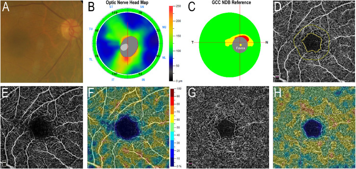
Figure 1.
Images of a 70-year-old woman with a high MMSE score (28) and normal DVP-VD. (A) Color fundus photo. (B) Optic nerve head map showing normal retinal nerve fiber layer thickness. (C) Ganglion cell complex layer was generally intact. (D) OCTA of full inner retinal layer showing the foveal avascular zone (inner yellowish line). (E) OCTA of SVP. (F) SVP-VD map. (G) OCTA of DVP. (H) DVP-VD map. The size of the foveal avascular zone and the percentage of parafoveal SVP-VD and parafoveal DVP-VD were 0.337 mm2, 52.3%, and 55.2%, respectively.

