Figure 6.
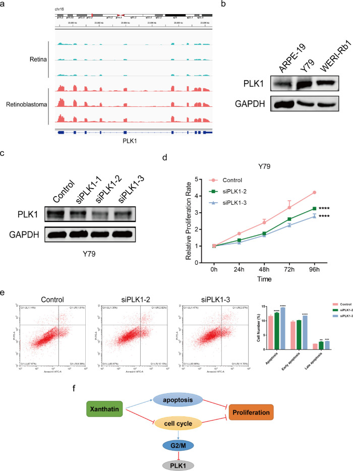
PLK1 contributed to tumor progression. (a) RNA-sequence analysis was performed to evaluate the transcriptome of three retinoblastoma samples and normal retina samples. (b) The expression level of PLK1 in the retinoblastoma cells was verified by Western blot. (c) Efficiency of PLK1 knockdown in Y79 cells by siRNAs was verified by Western blot. (d) CCK8 assay showing tumor cell growth after PLK1 knockdown. Cell growth was restrained in PLK1 KD Y79 cells. The absorbance values were detected at 0, 24, 48, 72, and 96 hours, and the control was arbitrarily set at 100% on 0 hour. ****P < 0.0001. (e) siPLK1 Y79 cells were more prone to apoptosis. The percentage of apoptotic cells was analyzed by flow cytometry. Error bars represent the SD of three replicates. The P values were from two-way ANOVA, **P < 0.01, ***P < 0.001, and ****P < 0.0001. (f) Schematic model of xanthatin inhibiting retinoblastoma cell growth via the PLK1-mediated G2/M cell cycle pathway.

