Figure 1.
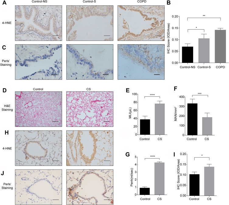
Ferroptosis is involved in human and murine COPD lung tissues. (A) Immunohistochemical (IHC) staining of 4-HNE in lung tissues from healthy volunteers (Control-NS), healthy smokers without COPD (Control-S) and COPD patients (original magnification ×400). Bar: 50 μm. The results were scored by (B) the integrated optical density (IOD)/area. (C) Fe3+ deposits were stained with Perls’ DAB in lung tissues from Control-NS, Control-S and COPD patients (original magnification ×400). Bar: 20 μm. (D–I) C57BL6 mice were exposed to room air (Control) or cigarette smoke (CS) for 6 months. (D) H&E staining in lung sections of mice in the control and CS groups (original magnification ×100). Bar: 200 μm. At least 3 areas in tissues collected from each mouse were captured, and 3 mice in each group were analyzed. (E and F) The alveolar size was measured by the mean linear intercept (MLI) and mean alveolar number (MAN) at ×100 magnification. (G) Enhanced pause (Penh) of room air- and CS-exposed mice. (H) IHC staining of 4-HNE from lung sections of room air- and CS-exposed mice (original magnification ×400). Bar: 50 μm. The results were scored by (I) the IOD/area. (J) Fe3+ deposits were stained with Perls’ DAB in air-exposed mice and CS-exposed mouse lungs (original magnification ×400). The results are presented as the means ± S.E.M. for three independent experiments. *P<0.05; **P < 0.01; ***P < 0.001; ****P < 0.0001.

