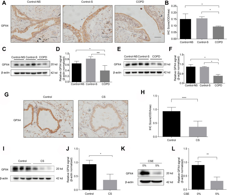
Figure 3.
GPX4 expression is downregulated in the lungs of COPD patients, murine models and CSE-treated HBE cells. (A) GPX4 immunostaining of lungs from Control-NS, Control-S and COPD patients is shown (original magnification ×400). Bar: 50 μm. The results were scored by (B) IOD/area. (CandD) Western blots of human lung homogenates from Control-NS, Control-S and COPD patients were probed using an anti-GPX4 antibody and normalized to β-actin levels (loading control). (E and F) Western blots of primary bronchial epithelial cells from Control-NS, Control-S and COPD patients were probed using an anti-GPX4 antibody and normalized to β-actin (loading control). (G–J) C57BL6 mice were exposed to room air (Control) or cigarette smoke (CS) for 6 months. (G and H) IHC staining of GPX4 in lungs from room air (Control)- and CS-exposed mice. Original magnification ×400. Bar: 50 μm. The results were scored by (H) IOD/area. (I and J) The expression levels of GPX4 in lung homogenates of room air (Control)- and CS-exposed mice were measured by Western blot analysis and normalized to those of β-actin. (K and L) The protein level of GPX4 in HBE cells treated with 0% or 5% CSE for 72 h was measured by Western blot and normalized to β-actin levels (loading control). The results are presented as the means ± S.E.M. from three independent experiments. *P<0.05; **P < 0.01; ***P < 0.001, ****P < 0.0001.

