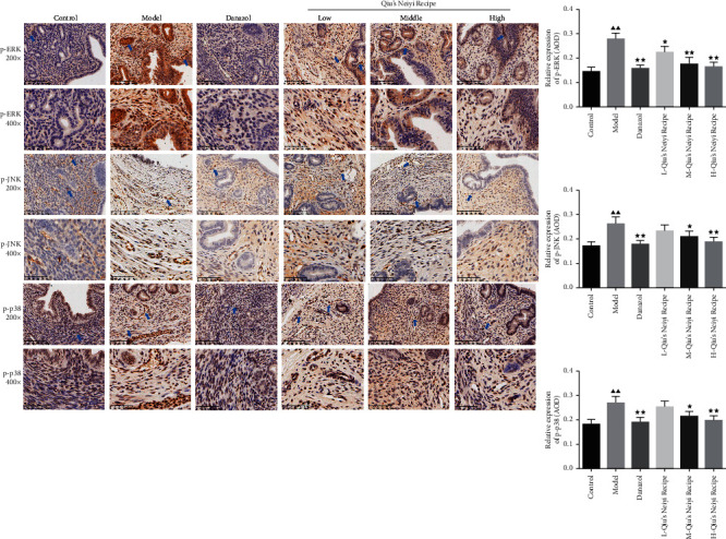
Figure 4.

Expressions of p-ERK, p-JNK, and p-p38 in uterine tissue were analyzed by immunochemical staining. (a) Representative microphotographs of immunochemical staining, original magnification, 200× and 400×. (b–d) Quantitative assessment of the expressions of p-ERK, p-JNK, and p-p38. L-Qiu's Neiyi recipe: a low dose of Qiu's Neiyi recipe; M-Qiu's Neiyi recipe: a middle dose of Qiu's Neiyi recipe; H-Qiu's Neiyi recipe: a high dose of Qiu's Neiyi recipe. Blue arrows indicate the target proteins in the immunochemical staining. , ΔΔP < 0.01 vs. control group, ∗P < 0.05, ∗∗P < 0.01 vs. model group.
