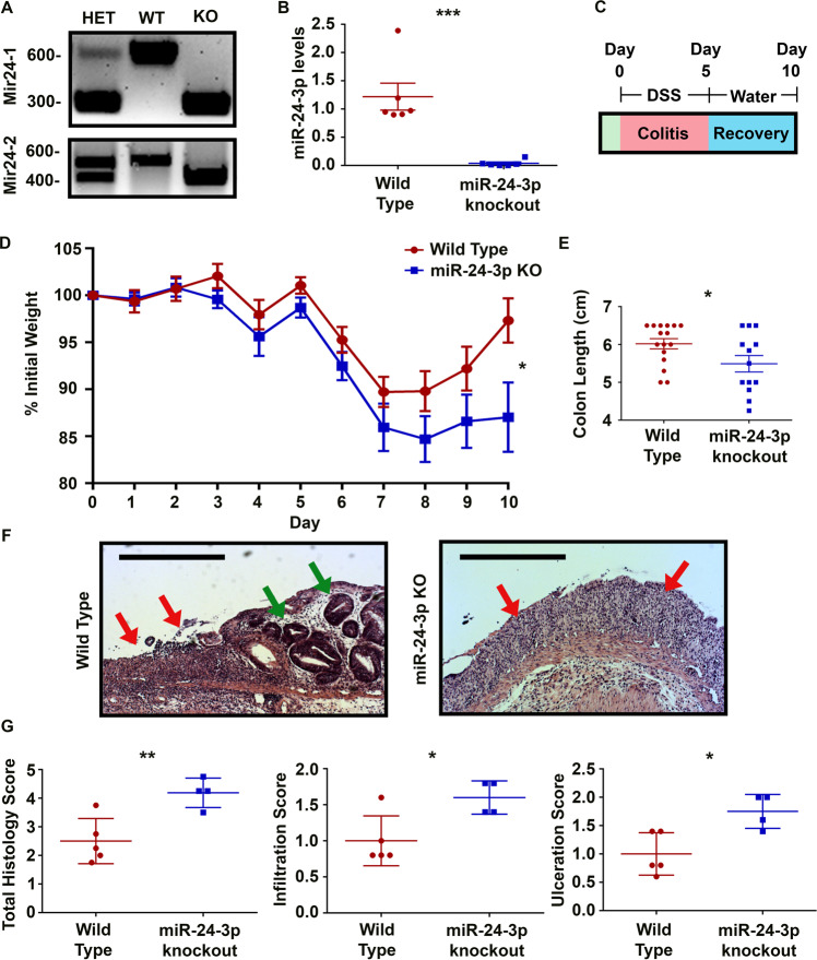
Fig. 1. miR-24-3p knockout mice exhibit reduced mucosal repair after colitis.
A Representative agarose gels of Mir24-1 and Mir24-2 genotyping. B After reverse transcription of colonic RNA, PCR was used to measure the level of miR-24-3p. n = 6 mice/group in two independent experiments. Mean ± SD. C A schematic describing the time course for colitis recovery experiments. D A graph of percent weight change over the course of recovery after colitis. n = 16/13 mice/group for wild type and miR-24-3p knockout, respectively, from three independent experiments. Mean ± SEM. E On day 10 of the protocol, mice were euthanized, and colon lengths were measured from the rectum to the cecum. n = 16/13 mice/group for wild type and miR-24-3p knockout, respectively, from three independent experiments. Mean ± SEM. F A representative image of the central colons of wild type or miR-24-3p knockout mice. Green arrows indicate areas of re-epithelization and red arrows indicate areas of ulceration. Scale bars = 0.5 mm. G Representative graphs of double-blind histology scores. n = 5/4 mice per group for wild type and miR-24-3p knockout, respectively, from two independent experiments. Mean ± SD. *p < 0.05; **p < 0.01; ***p < 0.001.

