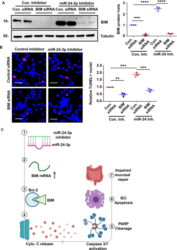
Fig. 8. Downregulation of BIM reduces the induction of apoptosis caused by miR-24-3p inhibition.
A Western blots and densitometric analysis of BIM and Tubulin from SW480 cells treated with staurosporine and either control or BIM siRNA plus either control inhibitor or miR-24-3p inhibitor. Three independent experiments. Mean ± SEM. B A TUNEL assay was used to measure the proportion of apoptotic cells. Magenta cells are TUNEL-positive and non-TUNEL-positive cell nuclei are labeled in blue. Three 250 × 250 µm fields were selected for quantification and are depicted in the graph. Three independent experiments. Mean ± SEM. Scale bars = 50 µm. **p < 0.01; ***p < 0.001; ****p < 0.0001. C We observed that when miR-24-3p was inhibited or removed from the genome (1) the mRNA and protein levels of BIM increase (2). BIM then stabilizes Bcl-2 (3) enabling Bax-mediated Cytochrome C release from the mitochondria (4). Cytochrome C activates caspases which result in events such as PARP cleavage (5). The activated pro-apoptotic enzymes then induce apoptosis in the intestinal epithelium (6). Induction of apoptosis during mucosal repair after colitis worsens outcomes (7).

