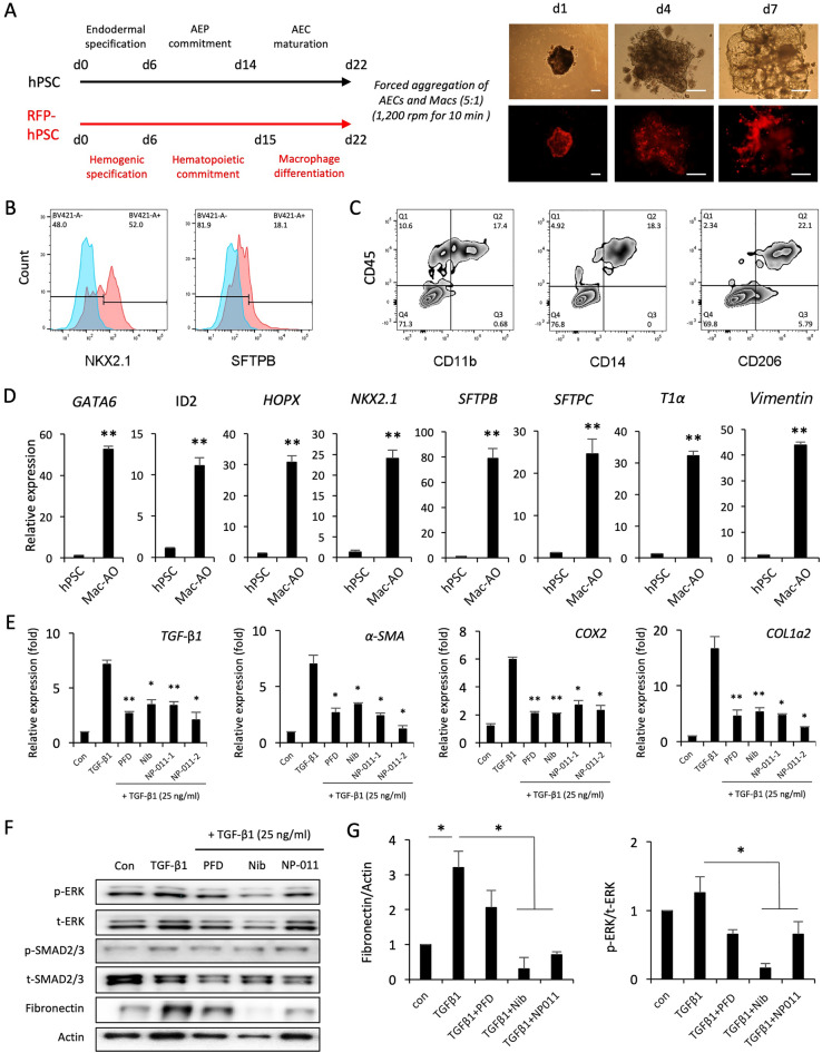
Fig. 2.
Comparison of anti-fibrotic effects of PFD, Nib, and NP-011 in TGF-β1-induced fibrotic Mac-AOs. A Schematic diagram of the generation of Mac-AOs from hPSCs with accompanying representative light and fluorescence microscopy images. Scale bars, 100 μm. B and C Flow cytometry analysis of the expression of AEC (NKX2.1 and SFTPB) and macrophage (CD11b, CD14 and CD206) markers on Mac-AOs. D qPCR of the indicated AEP (GATA6, HOPX, ID2, and NKX2.1), AEC1 (T1α), AEC2 (SFTPB and SFTPC), and mesenchymal stromal cell (Vimentin) markers in Mac-AOs. Data are shown as fold-change relative to undifferentiated hPSCs. E Effects of PFD (1 μg/mL), Nib (1 μM), and NP-011 (NP-011-1, 500 ng/mL; NP-011-2, 2 μg/mL) on the expression of fibrosis-related genes in TGF-β1-induced fibrotic Mac-AOs. F and G Western blotting and subsequent quantification of p-ERK, p-SMAD2/3, and Fibronectin in Mac-AOs from the indicated groups. Actin was used as a loading control. Data are presented as mean ± s.d. *p < 0.05, **p < 0.01

