FIGURE 1:
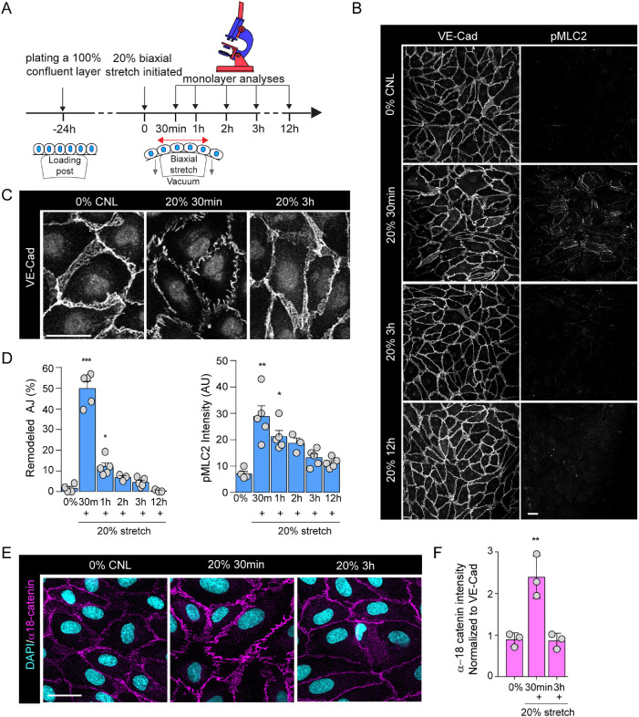
Cyclic mechanical stretch triggers transient adherens junction remodeling and actomyosin contraction. (A) Schematic illustration of stretch experiments. HUVECs were plated at confluency on silicon elastomers 24 h before stretch application, using negative pressure to deform the elastomer substrates (20%, 100 mHz), after which monolayers were analyzed at time points indicated. (B) Representative immunofluorescence images of VE-cadherin (VE-Cad) and pMLC2-stained HUVEC monolayers exposed to stretch. Note transient emergence of zipper-patterned adhesions and increased pMLC signal at 30 min of stretch. Scale bars 30 μm. (C) Close-up images of junctional rearrangements show reversibility of junctional zippering upon stretch. Scale bars 30 μm. (D) Quantification of AJ remodeling from VE-cadherin staining (left panel) and pMLC2 intensity (right panel). Mean ± SEM; n = 5 independent experiments; ***p = 0.0006, *p = 0.0203 (VE-cadherin) and) *p = 0.0168 and *p = 0.0071 (pMLC2), ANOVA, Dunnett’s. (E) Representative immunofluorescence images of α-18-stained HUVEC monolayers exposed to stretch. Note transient increase in α-18 intensity at 30 min of stretch. Scale bars 30 μm. (F) Quantification of α-18 catenin normalized to VE-cadherin intensity. Mean ± SD; n = 3 independent experiments; **p = 0.0018, ANOVA, Dunnett’s.

