FIGURE 4:
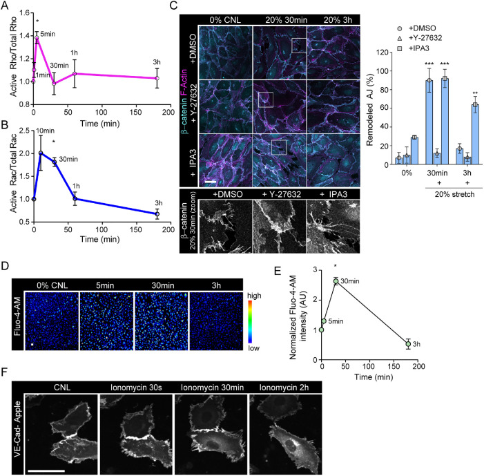
Stretch triggers transient calcium signaling and activation of Rho and GTPases to remodel junctions. (A) Quantification of RhoA activity over time shows peak of Rho activation at 5 min of stretch (mean ± SD; n = 3 independent experiments; *p = 0.0461, ANOVA with Dunnett’s). (B) Quantification of Rac1 activity over time shows peak of Rac activation at 30 min of stretch (mean ±SD; n = 4 independent experiments; *p = 0.0163, ANOVA with Dunnett’s). (C) Representative immunofluorescence images and quantification of AJ remodeling from β-catenin and F-actin (phalloidin)-stained HUVEC monolayers exposed to stretch in the presence of either Y-27632 or IPA3. Note prevention of adhesion zippering in Y-27632-treated cells and attenuated junction restoration in IPA3-treated cells. Scale bars 30 μm (mean ± SD; n = 3 independent experiments; ***p < 0.0001,**p = 0.0015, ANOVA with Dunnett’s). (D, E) Representative images, D, and quantification, E, of calcium imaging with Fluo-4-AM shows a transient increase in intracellular calcium at 30 min of stretch (mean ± SD; n = 3 independent experiments; *p = 0.0106, ANOVA with Dunnett’s; scale bars 30 μm). (F) Representative immunofluorescence images of confluent, transiently VE-cadherin-Apple transfected HUVEC monolayers showing remodeling of VE-cadherin junctions in response to ionophore application. Scale bars 30 μm.

