
FIGURE 4:
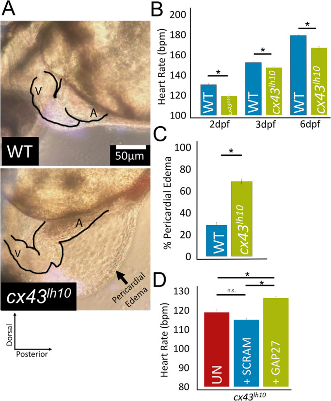
cx43lh10 zebrafish exhibit pronounced cardiac defects. (A) Representative images of the heart region of WT and cx43lh10 3 dpf embryos. cx43lh10 embryos exhibit malformed, underdeveloped, and elongated hearts, bradycardia, and pericardial edema. (B) Quantification of WT and cx43lh10 embryos at 2, 3, and 6 dpf indicates a significantly slower heart rate at all time points (n = 100 for WT and cx43lh10 embryos at 2, 3, and 6 dpf each, p ≤ 0.05). (C) Percentage of embryos exhibiting profound pericardial edema at 3 dpf (n = 496 for WT and 468 for cx43lh10 embryos, p ≤ 0.05). (D) Heart rates of uninjected 2 dpf cx43lh10 embryos (UN, red), 2 dpf cx43lh10 embryos injected with scrambled Gap27 peptide (SCRAM, blue), and 2 dpf cx43lh10 embryos injected with Gap27 peptide inhibitor (green). Note that the heart rate of embryos injected with Gap27 was restored close to WT levels (compare A with D) (n = 100 [uninfected], 21 [scrambled], 16 [Gap27 injected], p ≤ 0.05; error: SEM). (Also see Supplemental Movies 1 and 2.)